
We are the leading Preclinical Neuroscience CRO specializing in animal models of CNS diseases
We focus on providing research services leveraging the best available models of ALS, Alzheimer’s Disease, Tauopathies, Multiple Sclerosis, and Parkinson's Disease
Rodent Models
Learn more about our thoroughly characterized and validated animal models of neurological diseases
We have a range of transgenic and inducible mouse & rat models readily available for your studies. Our team of animal model scientists will work with you to find the right model.
CRO Services
We provide a range of specialized research services tailored to models of CNS diseases
End-to-end contract research services from model generation to in-life testing to quantitative tissue & fluid biomarkers.
- Animal Services
- Behavioral Testing
- Electrophysiology
- Fluid & Cell Biomarkers
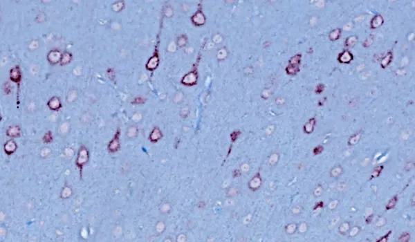
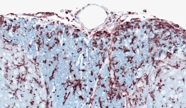
- Histology & IHC/mIF
- In Vivo Imaging
- Spatial Biology


Initiative
Microglia, Astrocytes, and Neurodegenerative Diseases
We explore the complex relationships between neuroinflammation and neurodegeneration, aiming to enhance scientific knowledge and accelerate the development of disease-modifying therapies.
By highlighting emerging scientific findings and reporting our original research, we seek to advance the understanding of how neuroinflammatory responses contribute to the onset and progression of neurodegenerative diseases, including ALS, Alzheimer's Disease & Tauopathies, and Parkinson's Disease.
Our Latest Innovations
Narrated presentations of our scientific advancements
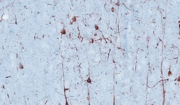
Astrocytes & Amyloid-β Mouse Models of Alzheimer's Disease
Analysis of astrocyte morphology in the amyloid-β plaque microenvironment provides a sensitive measure of disease progression in transgenic mice.
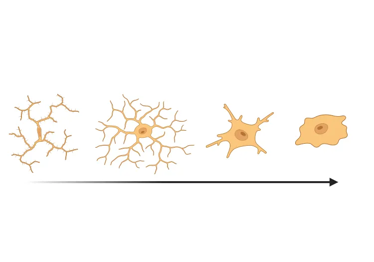
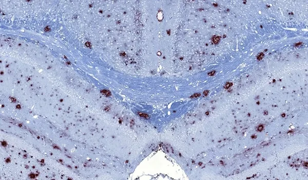
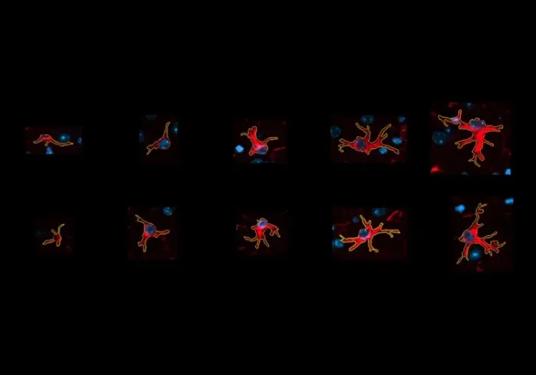
Microglial Activation in an α-Synuclein PFF Mouse Model
We have quantified microglial activation, based on morphology, in an α-synuclein preformed fibril (PFF) seeding & spreading mouse model of Parkinson’s disease.
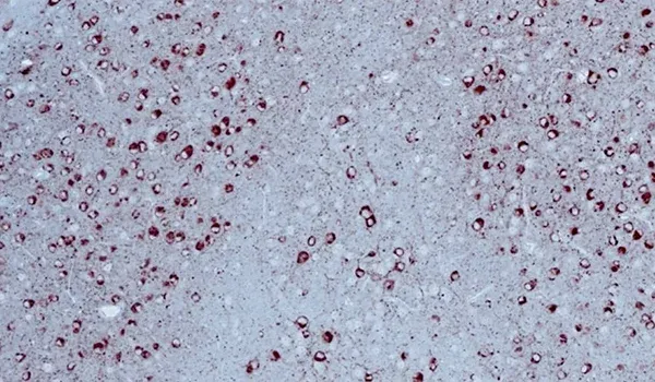
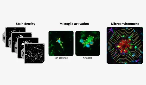
Amyloid-β & Inflammatory Microenvironment in Alzheimer's Mice
We have analyzed the complex spatial relationships between β-amyloid plaques, activated & resting microglia, and astrocytes in an APP/PS1 transgenic model.
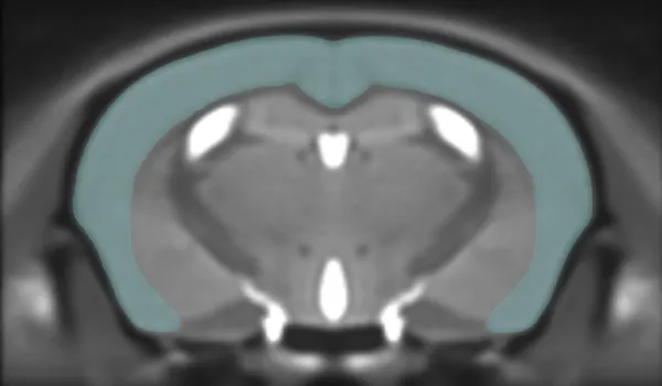
Brain Atrophy Analysis in Mouse Models of Neurodegeneration
Automated in vivo MRI-based quantitative brain atrophy measures (regional brain volumes and cortical thickness) from mouse models of ALS & Parkinson's disease.