Preclinical WebsiteClinical Website
Last Updated Date: September 05, 2024
Authors: Robin Guay-Lord and Barry J. Bedell, M.D., Ph.D.

Why is it important to characterize & quantify Aβ plaques?

Amyloid dysregulation is widely recognized as playing a central role in the progression of Alzheimer's disease (AD) (Selkoe, 2016). Hallmark neuropathologic features commonly seen in AD include extracellular amyloid-β (Aβ) plaques, astrogliosis, and microglial activation (Cohen, 2015). Amyloid plaques present a wide range of morphologies and neuroanatomical distributions. Both immunohistochemistry (IHC) and immunofluorescence (IF) can be employed to obtain detailed information about the localization, distribution, and morphology of Aβ plaques within specific brain regions, providing a level of detail not achievable with other quantification methods (e.g. brain imaging, fluid biomarkers).

Furthermore, clinical data suggests that studying only the global amyloid load in the brain may not give the full picture of the state of the pathology as multiple post-mortem studies have shown patients with abundant amyloid deposits at death without significant cognitive impairment (Katzman, 1988; Hulette, 1998; Aizenstein, 2008). Increasingly, scientists have attempted to link the presence of specific beta-amyloid plaque subtypes with clinical progression, and, for instance, have found that amyloid plaques lacking fibrillar structures (also called “diffuse plaques”) are often found in cases without cognitive impairment, in contrast to more compact fibrillar plaques that are more likely associated with cognitive impairment. The presence of swollen or dystrophic neurites in close proximity to the plaque has also been shown to be strongly correlated with cognitive impairment and disease severity (Dickson, 2001; Ly, 2011). These findings highlight the need for accurate characterization of the various amyloid plaque subtypes, which could help further our understanding of disease heterogeneity.

What are the various subtypes of amyloid-β plaques?

Amyloid-beta plaques originate from the cleavage of the amyloid precursor protein (APP) by the β- and γ-secretase enzymes. This process generates Aβ peptides, which can vary in length, with Aβ40 and Aβ42 being the most significant forms in AD. Aβ accumulation in the brain follows a specific spatiotemporal pattern, initially appearing in the neocortex and eventually spreading to subcortical regions, including the hippocampus (Braak, 1991; Braak, 2006). The Aβ peptide initially exists in a monomeric state, but can self-aggregate into various forms, such as short fibrillar oligomers, globular non-fibrillar oligomers, and amyloid fibrils that form aggregates known as plaques.

Spatiotemporal pattern of β-amyloid pathology progression (adapted from Braak, 1991).

Aβ plaques are classified, based on their morphology and fibril content, into diffuse and dense-core types using dyes that target the β-pleated sheet structure, such as Congo Red or Thioflavin-S (Serrano-Pozo, 2011). Thioflavin-S-negative diffuse plaques exhibit loose shapes and sizes, and lack a well-defined fibrillar structure. These diffuse plaques are commonly seen in the brains of cognitively-intact, elderly people. Thioflavin-S-positive dense-core plaques, on the other hand, have a compact core comprised of a cluster of extracellular filaments intermingled with surrounding neuronal, astrocytic, and microglial processes (Serrano-Pozo, 2011). These neuritic plaques (NPs) are associated with increased synaptic loss, neuron loss, astrogliosis, and microgliosis (Dickson, 2001; Ly, 2011; Tsering, 2023). NPs are commonly found in the later stages of AD, and have a higher correlation with cognitive decline than diffuse plaques (Haroutunian, 1998). This finding underscores the need to distinguish between diffuse and neuritic plaques when attempting to link β-amyloid burden and disease progression.

Aβ aggregates can also deposit in vessel walls, often referred to as vascular Aβ or cerebral amyloid angiopathy (CAA). These deposits are usually formed from Aβ40 peptides, which are more soluble than the Aβ42 peptides that are commonly found in typical parenchymal plaques. Studies have shown that CAA can be a contributing factor to cognitive decline seen in AD (Greenberg, 2004; Arvanitakis, 2011).

Illustration of the various types of amyloid-β plaques.

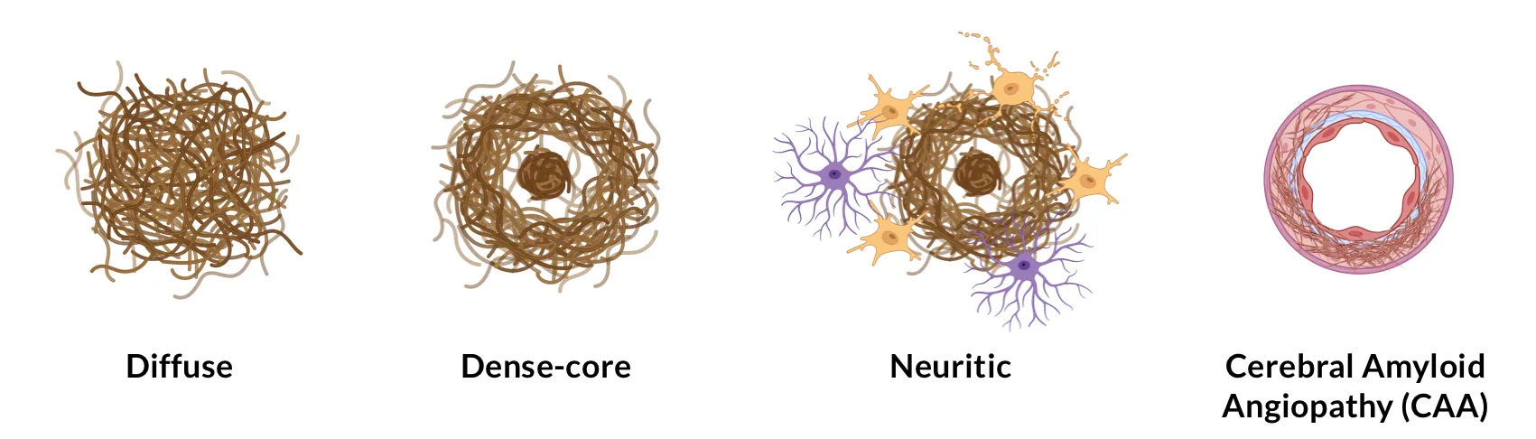
Illustration of various types of amyloid-β plaques.

Other rarer subtypes of Aβ plaques include dense-core coarse-grained plaques, which are associated with the early stages of AD (Boon, 2020), and diffuse cotton wool plaques, often found in patients with specific genetic mutations, such as those in the PSEN1 gene (Crook, 1998).

How are amyloid plaques characterized in IHC and multiplex IF images?

The assessment of plaque burden in immunohistochemistry (IHC) and immunofluorescence (IF) data is conventionally preformed manually using semi-quantitative criteria established by the Consortium to Establish a Registry for Alzheimer’s Disease (CERAD) (Mirra, 1991). These criteria focus on assessing the highest density of neocortical neuritic plaques, and they do not consider the presence of diffuse plaques in the scoring calculation.

Significant efforts have been made to develop quantitative approaches to assess Aβ plaque load that account for anatomical location, plaque density, and plaque subtype. Common methods employed to quantify these features manually include unbiased stereological counts of plaques (Busch, 1997; Ohm, 1997), and grading of plaque densities using a numerical rating scale (Arnold, 1991). These manual assessments can be tedious, prone to interrater variability, and hard to scale for larger studies. Automated methods that rely on classical computer vision algorithms (e.g. thresholding, morphological operations, etc.) have been investigated to reduce the burden on pathologists and increase the reliability of the quantification (Byrne, 2009; Neltner, 2012; Samaroo, 2012; Kapasi, 2023). However, a vast majority of these classical methods still require human-defined inputs, making them susceptible to batch variations and staining variability observed in larger datasets.

Our group has performed quantification of amyloid plaques in a mouse model with early-onset and progressive AD-like accumulation of β-amyloid deposits. This transgenic mouse line (ARTE10) is characterized by overexpression of mutated APP and PS1 (Willuweit, 2009). Aβ plaque load was assessed from IF images of these mice at different timepoints using a combination of blurring, local hysteresis thresholding, and morphological operations. This approach was fully-automated and did not require any human-defined inputs to quantify Aβ load in tissue sections. See our Presentation – Amyloid-β & Inflammatory Microenvironment in Alzheimer’s Mice.

Microscopy Images

The Interactive Image Viewer below allows you to explore the entire multiplex immunofluorescence tissue section.

You can pan around the image using the left mouse button. You can zoom in & out using the mouse/trackpad (up/down) or the + and - buttons in the upper left corner. You can toggle (on/off), change color, and adjust image settings for the channels and segmentations in the Control Panel in the upper right corner.


In this visualization, an APP/PS1 (ARTE10) mouse brain is shown with the "Aβ Plaques" Segmentation Masks (yellow), which can be checked/unchecked to hide/show the underlying "Amyloid-β" staining (red).

Control Panel
Section: APP/PS1 Mouse
Segmentations
Channels

Multiplex immunofluorescence stained brain tissue section that demonstrates Aβ plaques (along with segmentation), activated microglia, and reactive astrocytes in a 12 month-old APP/PS1 (ARTE10) transgenic mouse. Note that the sensitivity of the Aβ plaque segmentation can be modified during image processing via various parameter settings and morphological operations.

More recently, research groups have reported using deep-learning approaches for automated classification of AD plaques into plaque subtypes, leveraging the expert-level performance of these models to recognize intricate patterns in images. For example, Tang and colleagues (Tang, 2019) developed a proof-of-concept deep learning pipeline that analyzes whole-slide images, and classifies Aβ pathologies between diffuse plaques, dense-core plaques, and CAA. The same group later expanded on those results by showing that these machine learning models were robust to cohort variations across multi-center studies (Vizcarra, 2020; Wong, 2022). Other groups have reported using similar approaches to differentiate tauopathies (Signaevsky, 2019; Koga, 2022; Wong, 2022). Overall, extensive efforts have been made to train these deep-learning models on a wide range of input parameters (brain region, staining methodology, pathological features) to make them adaptable and generalizable.

Our team would be happy to answer any questions about amyloid-β plaque analysis or provide specific information about the Alzheimer’s disease models that we use for therapeutic efficacy studies.

Discover more about our Alzheimer's Disease Models

FAQs

Which markers are commonly used to identify amyloid plaques?

Typically, plaques are identified and classified with standard histology using dyes, such as Congo Red, Thioflavin-S, or Gallyas silver stain. Immunohistochemistry (IHC) and immunofluorescence (IF) methods can use anti-Aβ antibodies or anti-amyloid fibril (OC) antibodies to stain the total amyloid and use thioflavin-S to discriminate between diffuse plaques and core plaques. Additionally, other fluorescent channels can be used to highlight the various glial cells (astrocytes, microglia) present in the microenvironment of the amyloid plaques.


What are some other methods to quantify amyloid burden in the context of AD?

Overall amyloid burden can be measured in vivo using non-invasive imaging techniques, such as positron emission tomography (PET). It can also be measured via various biofluid (e.g. blood, CSF) markers using a wide range of ‘omics techniques. While these methods provide valuable longitudinal readouts, they lack the spatial resolution and do not offer information on the cellular environment of the amyloid plaques.


How is amyloid quantified in commonly used mouse models of AD?

Extensive work has been performed over the years to quantify the spatiotemporal accumulation of  Aβ in a wide variety of mouse models of AD, including ,but not limited to, APP/PS1 mice (Ferguson, 2013; Lok, 2013; Sasaguri, 2017; Willuweit, 2009), 5xFAD mice (Forner, 2021; Liu, 2017; Oh, 2018), and Tg2576 mice (Han, 2012; Liu, 2017). A wide variety of quantification methods were used for this purpose, ranging from manual stereological identification to fully-automated quantification algorithms.


What are the main challenges encountered when quantifying amyloid plaques in transgenic rodent models of Alzheimer's disease?

Quantifying amyloid plaques in rodent models presents challenges related to variability in plaque distribution, staining intensity, and background noise in IHC and IF images. IHC images are also subject to folds, tears, and artifacts that can interfere with the accurate quantification of amyloid burden. Developing standardized protocols for optimized tissue processing, antibody dilutions, staining protocols, and image acquisition parameters can help minimize variability and ensure reproducibility across studies. Collaborative efforts, such as inter-laboratory comparisons and proficiency testing, can also help to establish consensus protocols and guidelines for accurate plaque quantification and data interpretation.


References

Aizenstein, H. J., Nebes, R. D., Saxton, J. A., Price, J. C., Mathis, C. A., Tsopelas, N. D., Ziolko, S. K., James, J. A., Snitz, B. E., Houck, P. R., Bi, W., Cohen, A. D., Lopresti, B. J., DeKosky, S. T., Halligan, E. M., Klunk, W. E. Frequent amyloid deposition without significant cognitive impairment among the elderly. Arch. Neurol., 65: 1509-1517, 2008; doi: 10.1001/archneur.65.11.1509

Arnold, S. E., Hyman, B. T., Flory, J., Damasio, A. R., Van Hoesen, G. W. The topographical and neuroanatomical distribution of neurofibrillary tangles and neuritic plaques in the cerebral cortex of patients with Alzheimer’s disease. Cereb. Cortex, 1: 103–116, 1991; doi: 10.1093/cercor/1.1.103

Arvanitakis, Z., Leurgans, S. E., Wang, Z., Wilson, R. S., Bennett, D. A., Schneider, J. A. Cerebral amyloid angiopathy pathology and cognitive domains in older persons. Ann. Neurol., 69: 320–327, 2011; doi: 10.1002/ana.22112

Braak, H., Braak, E. Neuropathological stageing of Alzheimer-related changes. Acta Neuropathol., 82: 239–259, 1991; doi: 10.1007/bf00308809

Braak, H., Alafuzoff, I., Arzberger, T., Kretzschmar, H., Tredici, K. Staging of Alzheimer disease-associated neurofibrillary pathology using paraffin sections and immunocytochemistry. Acta Neuropathol., 112: 389, 2006; doi: 10.1007/s00401-006-0127-z

Boon, B. D. C., Bulk, M., Jonker, A. J., Morrema, T. H. J., van den Berg, E., Popovic, M., Walter, J., Kumar, S., van der Lee, S. J., Holstege, H., Zhu, X., Van Nostrand, W. E., Natté, R., van der Weerd, L., Bouwman, F. H., van de Berg, W. D. J., Rozemuller, A. J. M., Hoozemans, J. J. M. The coarse-grained plaque: a divergent Aβ plaque-type in early-onset Alzheimer’s disease. Acta Neuropathol., 140: 811–830, 2020; doi: 10.1007/s00401-020-02198-8

Busch, C., Bohl, J., Ohm, T. G. Spatial, temporal and numeric analysis of Alzheimer changes in the nucleus coeruleus. Neurobiol. Aging, 18: 401–406, 1997; doi: 10.1016/s0197-4580(97)00035-3

Byrne, U. T. E., Ross, J. M., Faull, R. L. M., Dragunow, M. High-throughput quantification of Alzheimer’s disease pathological markers in the post-mortem human brain. J. Neurosci. Methods, 176: 298–309, 2009; doi: 10.1016/j.jneumeth.2008.09.008

Cohen, M. L., Kim, C., Haldiman, T., ElHag, M., Mehndiratta, P., Pichet, T., Lissemore, F., Shea, M., Cohen, Y., Chen, W., Blevins, J., Appleby, B. S., Surewicz, K., Surewicz, W. K., Sajatovic, M., Tatsuoka, C., Zhang, S., Mayo, P., … Safar, J. G. Rapidly progressive Alzheimer’s disease features distinct structures of amyloid-β. Brain,138: 1009–1022, 2015; doi: 10.1093/brain/awv006

Crook, R., Verkkoniemi, A., Perez-Tur, J., Mehta, N., Baker, M., Houlden, H., Farrer, M., Hutton, M., Lincoln, S., Hardy, J., Gwinn, K., Somer, M., Paetau, A., Kalimo, H., Ylikoski, R., Pöyhönen, M., Kucera, S., Haltia, M. A variant of Alzheimer’s disease with spastic paraparesis and unusual plaques due to deletion of exon 9 of presenilin 1. Nat. Med., 4: 452–455, 1998; doi: 10.1038/nm0498-452

Dickson, T. C., Vickers, J. C. The morphological phenotype of β-amyloid plaques and associated neuritic changes in Alzheimer’s disease. Neuroscience, 105: 99–107, 2001; doi: 10.1016/s0306-4522(01)00169-5

Ferguson, S. A., Sarkar, S., Schmued, L. C. Longitudinal behavioral changes in the APP/PS1 transgenic Alzheimer’s Disease model. Behav. Brain Res., 242: 125–134, 2013: doi: 10.1016/j.bbr.2012.12.055

Forner, S., Kawauchi, S., Balderrama-Gutierrez, G., Kramár, E. A., Matheos, D. P., Phan, J., Javonillo, D. I., Tran, K. M., Hingco, E., da Cunha, C., Rezaie, N., Alcantara, J. A., Baglietto-Vargas, D., Jansen, C., Neumann, J., Wood, M. A., MacGregor, G. R., Mortazavi, A., … Green, K. N. Systematic phenotyping and characterization of the 5xFAD mouse model of Alzheimer’s disease. Sci. Data, 8: 1–16, 2021; doi: 10.1038/s41597-021-01054-y

Greenberg, S. M., Gurol, M. E., Rosand, J., Smith, E. E. Amyloid angiopathy-related vascular cognitive impairment. Stroke, 35: 2616–2619, 2004; doi: 10.1161/01.STR.0000143224.36527.44

Han, P. L., Kim, T. K., Lee, J. E., Park, S. K., Lee, K. W., Seo, J. S., Im, J. Y., Kim, S. T., Lee, J. Y., Kim, Y. H., Lee, J. K. Analysis of differential plaque depositions in the brains of Tg2576 and Tg-APPswe/PS1dE9 transgenic mouse models of Alzheimer disease. Exp. Mol. Med., 44: 492–502, 2012; doi: 10.3858/emm.2012.44.8.056

Haroutunian, V., Perl, D. P., Purohit, D. P., Marin, D., Khan, K., Lantz, M., Davis, K. L., Mohs, R. C. Regional distribution of neuritic plaques in the nondemented elderly and subjects with very mild Alzheimer disease. Arch. Neurol., 55: 1185–1191, 1998; doi: 10.1001/archneur.55.9.1185

Hulette, C. M., Welsh-Bohmer, K. A., Murray, M. G., Saunders, A. M., Mash, D. C., McIntyre, L. M. Neuropathological and neuropsychological changes in “normal” aging: evidence for preclinical Alzheimer disease in cognitively normal individuals. J. Neuropathol. Exp. Neurol., 57: 1168–1174, 1998; doi: 10.1097/00005072-199812000-00009

Kapasi, A., Poirier, J., Hedayat, A., Scherlek, A., Mondal, S., Wu, T., Gibbons, J., Barnes, L. L., Bennett, D. A., Leurgans, S. E., Schneider, J. A. High-throughput digital quantification of Alzheimer disease pathology and associated infrastructure in large autopsy studies. J. Neuropathol. Exp. Neurol., 82: 976, 2023; doi: 10.1093/jnen/nlad086

Katzman, R., Terry, R., DeTeresa, R., Brown, T., Davies, P., Fuld, P., Renbing, X., Peck, A. Clinical, pathological, and neurochemical changes in dementia: A subgroup with preserved mental status and numerous neocortical plaques. Ann. Neurol., 23: 138–144, 1988; doi: 10.1002/ana.410230206

Koga, S., Ikeda, A., Dickson, D. W. Deep learning‐based model for diagnosing Alzheimer’s disease and tauopathies. Neuropathol. Appl. Neurobiol., 48: e12759, 2022; doi: 10.1111/nan.12759

Liu, P., Reichl, J. H., Rao, E. R., McNellis, B. M., Huang, E. S., Hemmy, L. S., Forster, C. L., Kuskowski, M. A., Borchelt, D. R., Vassar, R., Ashe, K. H., Zahs, K. R. Quantitative comparison of dense-core amyloid plaque accumulation in amyloid-β precursor protein transgenic mice. J. Alzheimers Dis., 56: 743, 2017: doi: 10.3233/JAD-161027

Lok, K., Zhao, H., Shen, H., Wang, Z., Gao, X., Zhao, W., Yin, M. Characterization of the APP/PS1 mouse model of Alzheimer’s disease in senescence accelerated background. Neurosci. Lett., 557: 84–89, 2013; doi: 10.1016/j.neulet.2013.10.051

Ly, P. T. T., Cai, F., Song, W. Detection of neuritic plaques in Alzheimer’s disease mouse model. J. Vis. Exp., 53: e2831, 2011; doi: 10.3791/2831

Mirra, S. S., Heyman, A., McKeel, D., Sumi, S. M., Crain, B. J., Brownlee, L. M., Vogel, F. S., Hughes, J. P., van Belle, G., Berg, L., Ball, M. J., Bierer, L. M., Claasen, D., Hansen, L. R., Hart, M., Hedreen, J., Baltimore, B., Hen Derson, V., … Terry, R. D. The Consortium to Establish a Registry for Alzheimer’s Disease (CERAD). Part II. Standardization of the neuropathologic assessment of Alzheimer’s disease. Neurology, 41: 479–486, 1991; doi: 10.1212/wnl.41.4.479

Neltner, J. H., Abner, E. L., Schmitt, F. A., Denison, S. K., Anderson, S., Patel, E., Nelson, P. T. Digital pathology and image analysis for robust high-throughput quantitative assessment of Alzheimer disease neuropathologic changes. J. Neuropathol. Exp. Neurol., 71: 1075-1085, 2012; doi: 10.1097/NEN.0b013e3182768de4

Oh, S. J., Lee, H. J., Kang, K. J., Han, S. J., Lee, Y. J., Lee, K. C., Lim, S. M., Chi, D. Y., Kim, K. M., Park, J. A., Choi, J. Y. Early detection of Aβ deposition in the 5xFAD mouse by amyloid PET. Contrast Media Mol. Imaging, 2018: 5272014, 2018; doi: 10.1155/2018/5272014

Ohm, T. G., Busch, C., Bohl, J. Unbiased estimation of neuronal numbers in the human nucleus coeruleus during aging. Neurobiol. Aging,18: 393–399, 1997; doi: 10.1016/s0197-4580(97)00034-1

Samaroo, H. D., Opsahl, A. C., Schreiber, J., O’Neill, S. M., Marconi, M., Qian, J., Carvajal-Gonzalez, S., Tate, B., Milici, A. J., Bales, K. R., Stephenson, D. T. High throughput object-based image analysis of β-amyloid plaques in human and transgenic mouse brain. J. Neurosci. Methods, 204: 179–188, 2012; doi: 10.1016/j.jneumeth.2011.10.003

Sasaguri, H., Nilsson, P., Hashimoto, S., Nagata, K., Saito, T., Strooper, B. De, Hardy, J., Vassar, R., Winblad, B., Saido, T. C. APP mouse models for Alzheimer’s disease preclinical studies. EMBO J., 36: 2473, 2017; doi: 10.15252/embj.201797397

Selkoe, D. J., Hardy, J. The amyloid hypothesis of Alzheimer’s disease at 25 years. EMBO Mol. Med., 8: 595-608, 2016; doi: 10.15252/emmm.201606210

Serrano-Pozo, A., Frosch, M. P., Masliah, E., Hyman, B. T. Neuropathological alterations in Alzheimer disease. Cold Spring Harb. Perspect. Med., 1: a006189, 2011; doi: 10.1101/cshperspect.a006189

Signaevsky, M., Prastawa, M., Farrell, K., Tabish, N., Baldwin, E., Han, N., Iida, M. A., Koll, J., Bryce, C., Purohit, D., Haroutunian, V., McKee, A. C., Stein, T. D., White, C. L., Walker, J., Richardson, T. E., Hanson, R., Donovan, M. J., … Crary, J. F. Artificial intelligence in neuropathology: deep learning-based assessment of tauopathy. Lab. Invest., 99: 1019–1029, 2019; doi: 10.1038/s41374-019-0202-4

Tang, Z., Chuang, K. V., DeCarli, C., Jin, L. W., Beckett, L., Keiser, M. J., Dugger, B. N. Interpretable classification of Alzheimer’s disease pathologies with a convolutional neural network pipeline. Nature Commun.,10: 2173, 2019; doi: 10.1038/s41467-019-10212-1

Tsering, W., Prokop, S. Neuritic plaques — gateways to understanding Alzheimer’s disease. Molecular Neurobiol., 61: 2808–2821, 2023; doi: 10.1007/s12035-023-03736-7

Vizcarra, J. C., Gearing, M., Keiser, M. J., Glass, J. D., Dugger, B. N., Gutman, D. A. Validation of machine learning models to detect amyloid pathologies across institutions. Acta Neuropathol. Commun., 8: 59, 2020; doi: 10.1186/s40478-020-00927-4

Willuweit, A., Velden, J., Godemann, R., Manook, A., Jetzek, F., Tintrup, H., Kauselmann, G., Zevnik, B., Henriksen, G., Drzezga, A., Pohlner, J., Schoor, M., Kemp, J. A., von der Kammer, H. Early-onset and robust amyloid pathology in a new homozygous mouse model of Alzheimer’s disease. PLoS One, 4: e7931, 2009; doi: 10.1371/journal.pone.0007931

Wong, D. R., Tang, Z., Mew, N. C., Das, S., Athey, J., McAleese, K. E., Kofler, J. K., Flanagan, M. E., Borys, E., White, C. L., Butte, A. J., Dugger, B. N., Keiser, M. J. Deep learning from multiple experts improves identification of amyloid neuropathologies. Acta. Neuropathol. Commun., 10: 66, 2022; doi: 10.1186/s40478-022-01365-0


Keywords

Alzheimer's Disease: a progressive neurodegenerative disorder characterized by cognitive decline, memory loss, and behavioral changes. It is associated with the accumulation of amyloid-beta plaques and tau tangles in the brain, and loss of neurons and synapses in the cerebral cortex and subcortical regions.  

Amyloid-beta (Aβ): A peptide derived from the amyloid precursor protein (APP) that can aggregate to form plaques.

Astrogliosis: The proliferation and hypertrophy of astrocytes, a type of glial cell, in response to brain injury or disease, often observed in neurodegenerative diseases.

Cerebral Amyloid Angiopathy (CAA): A condition where amyloid-beta plaques accumulate in the walls of cerebral blood vessels, potentially contributing to cognitive decline and increasing the risk of hemorrhagic stroke.

Cognitive Impairment: A decline in cognitive function, including memory, thinking, and reasoning skills.

Deep Learning: A subset of machine learning techniques that use neural networks with many layers (deep neural networks) to analyze complex patterns in data.

Diffuse Plaques: A subtype of amyloid-beta plaques that lack a compact fibrillar structure and are often associated with cases without cognitive impairment.

Immunofluorescence (IF): A method similar to immunohistochemistry that uses fluorescently-labeled antibodies to detect specific antigens in tissue samples.

Immunohistochemistry (IHC): a laboratory technique to rapidly identify specific proteins in cells and tissues. IHC capitalizes on the ability of antibodies to target specific proteins, and then utilizes a sandwich of secondary antibodies and detection reagents to identify and localize the protein of interest in tissue sections at the microscopic level. 

Neuritic Plaques: Amyloid plaque containing dystrophic neurites (degenerating axons and dendrites).

Neurodegeneration: a complex, multifactorial process resulting in the loss of neurons.

Omics Techniques: A range of methodologies used to study biological molecules on a large scale, such as genomics, proteomics, and metabolomics.


Related Content

Up-to-date information on Alzheimer's Disease and best practices related to the evaluation of therapeutic agents in AD animal models.

More Information

Let us know what you’re interested in. Our team will be happy to discuss with you!

Email us at i[email protected] or simply complete & submit the form below. 

Name*
Email*
Phone Number*
Affiliation (Company/University)*
Message*

Your privacy is important to us. We will protect your data as outlined in our Privacy Notice.

I agree to the terms in the Privacy Notice*

We use necessary cookies to make our site work. We also use other cookies to help us make improvements by measuring how you use the site or for marketing purposes. You have the choice to accept or reject them all. For more detailed information about the cookies we use, see our Privacy Notice.