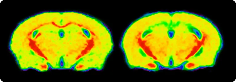
Biospective’s cuprizone mouse model facilitates multiple sclerosis (MS) drug development with translational MS pathology for preclinical research. As a neuroscience CRO with extensive experience with cuprizone models, we offer comprehensive in vivo services — including therapeutic efficacy, mechanism-of-action, and target engagement — supported by clinically relevant biomarkers, including advanced MRI imaging (e.g. MTR) and quantitative multiplex immunofluorescence (mIF).
The cuprizone mouse model is considered a gold-standard model for multiple sclerosis drug development studies. This mouse model reliably demonstrates toxin-driven demyelination, remyelination, OPC proliferation, migration & maturation, and neuroinflammation reflecting key pathologic features of human MS. At Biospective, we have rigorously validated translational in vivo MRI measures and quantitative IHC & multiplex immunofluorescence markers (e.g. MBP, oligodendrocyte lineage markers, Iba1, GFAP) for this MS model to generate pharma-grade data for our sponsors around the world.
Overview of the Cuprizone Mouse Model of MS
A gold-standard animal model of MS for preclinical drug development.
The cuprizone model is a widely-used animal model for assessing therapeutic agents targeting demyelination of the central nervous system (CNS). Cuprizone induction is most commonly performed by feeding mice with:
- 0.2-0.3% cuprizone
- 0.2-0.3% cuprizone + rapamycin
Mice are typically fed the copper-chelating cuprizone toxin for 5-6 weeks and then normal diet is restored allowing for assessment of the ability of a therapeutic to accelerate remyelination or modulate neuroinflammation. The addition of rapamycin to the standard cuprizone protocol results in slower, incomplete disease resolution following the withdrawal of the cuprizone diet.
Cuprizone mice model several key aspects of human MS, including the following key pathologic features:
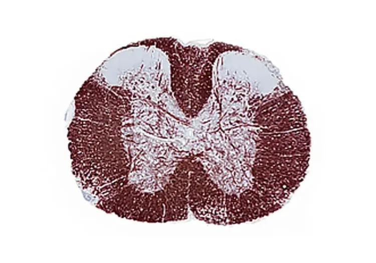
- Demyelination: Extensive loss of myelin (e.g. MBP staining) is observed in the corpus callosum.
-
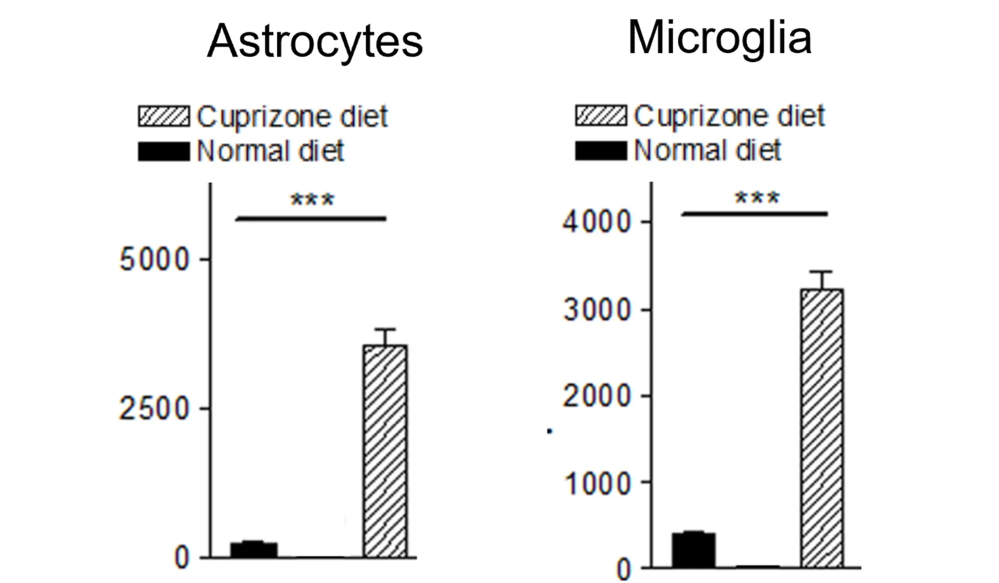
Neuroinflammation: Activated microglia and reactive astrocytes are present in the absence of peripheral inflammatory infiltrates.
- OPC proliferation, migration & maturation: Oligodendrocyte Precursor Cells (OPCs) are active in the cuprizone model and are responsible for the remyelination phase of this disease model.
The evolution and resolution of pathology in the cuprizone model is highly predictable, which makes it an attractive model for the evaluation of therapeutics. Unlike EAE, the cuprizone model of multiple sclerosis is not autoimmune mediated, and the cuprizone induced white matter demyelination and remyelination are not confounded by a peripheral immune response. EAE and cuprizone mice model different aspects of MS pathology, and therapeutic studies are often performed in parallel. Learn more in our Resource - Demyelination & Remyelination in the Cuprizone Model.
Validated Endpoints & Translational Biomarkers
Biospective has implemented a suite of validated endpoints and MS relevant biomarkers to enable clinical advancement of therapeutic programs.
To fully characterize cuprizone-fed mice and assess treatment outcomes, Biospective has validated a broad spectrum of endpoints, including imaging biomarkers and histopathology. This comprehensive approach yields robust, quantitative readouts for both efficacy and mechanism-of-action in preclinical studies. Key validated endpoints in our cuprizone mouse model include:
-
In vivo MRI: By using Magnetization Transfer Imaging (MTI), we are able to obtain a sensitive, non-invasive measure of disease progression in the corpus callosum.
- Quantitative Histopathology (IHC/mIF) of Brain: High-resolution tissue analyses to quantify MS-related pathology. We perform immunohistochemistry (IHC) and multiplex immunofluorescence for markers such as myelin, oligodendrocyte precursor cells (OPCs), mature oligodendrocytes, microglia, and astrocytes. Digital image analysis of these stained tissues provides quantitative measures of demyelination and inflammation in the corpus callosum.
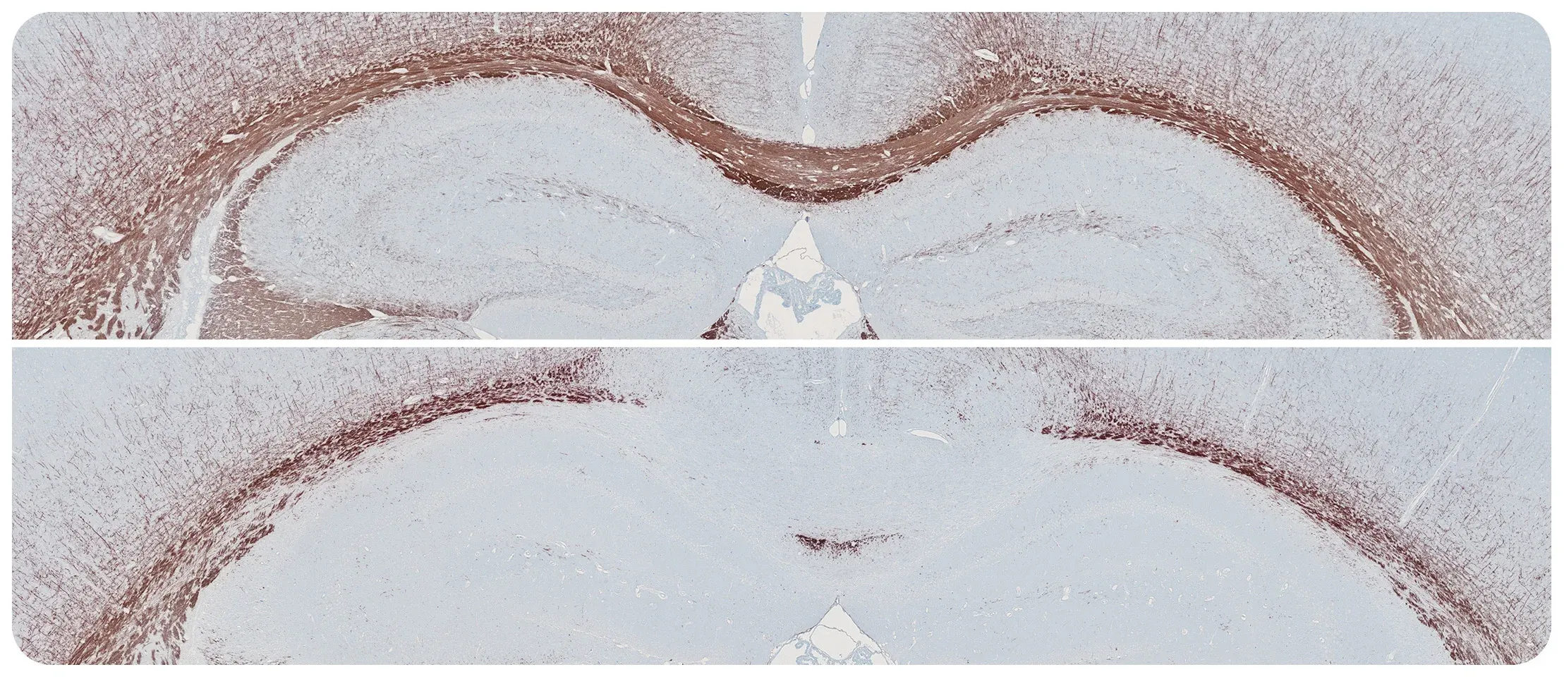
Myelin Basic Protein (MBP) immunohistochemistry (IHC) staining of the corpus callosum in control mice (top) and mice after 5 weeks of cuprizone diet treatment (bottom).
Biospective's Cuprizone Model Expertise and Services
Biospective is a global neuroscience CRO with deep expertise in MS animal models, including the cuprizone model, which is a core part of our service portfolio.
Our team has over 15 years of experience executing studies in cuprizone models. We bring our scientific and operational expertise to provide high-quality studies to our sponsors seeking to outsource their in vivo research.
Some key advantages of partnering with Biospective for cuprizone model studies studies include:
-
Extensive Experience & Model Characterization: We have extensively characterized the cuprizone mouse model through numerous studies over many years, generating datasets that inform best practices and enhance reproducibility. This track record underscores our unique expertise with this MS model.
-
End-to-End Preclinical Services: Biospective provides integrated services from study design through execution and data analysis. Our capabilities include comprehensive in-life assessments (behavioral testing, motor function assays, etc.), neuroimaging (MRI, PET, SPECT, CT), bioanalysis (fluid biomarkers), IHC & multiplex immunofluorescence), and expert data interpretation. This one-stop approach ensures consistency and accelerates timelines.
-
Translational Biomarkers & Readouts: We incorporate translational endpoints that bridge preclinical findings to clinical outcomes. For example, we perform in vivo imaging (MRI) in cuprizone mice to longitudinally follow disease evolution and resolution, analogous to what is performed in patients. These biomarkers enhance the translatability of study results to human trials.
-
Global Collaboration & Flexibility: We are a global preclinical neuroscience CRO serving biotech and pharmaceutical clients worldwide. Our scientists collaborate closely with sponsors to tailor studies to specific therapeutic mechanisms or targets. We can accommodate custom endpoints or novel treatment paradigms. We also offer flexibility in study design to meet your program’s needs. Importantly, we prioritize scientific rigor, reproducibility, and open communication throughout the partnership.
By leveraging these strengths, Biospective enables biotech and pharma teams to generate decision-quality data in the cuprizone mouse model efficiently. We pride ourselves on fast project initiation, clear data reporting, and supporting our clients across the preclinical phases of drug development.
Leverage Biospective's Cuprizone Model for MS Drug Development
By partnering with Biospective for your MS research, you gain access to an internationally recognized team of neurobiology experts and a deeply characterized preclinical model that can accelerate your drug development pipeline.
We have extensive experience executing studies in the cuprizone mouse model across a range of therapeutic modalities (small molecules, biologics, antibodies, gene therapies, antisense oligonucleotides, etc.). At Biospective, we have the ability to effectively handle large-scale studies and we provide seamless end-to-end integration of all study components. We handle every aspect of the experiment, including model induction, longitudinal in vivo imaging, biofluid collection/processing, and post-mortem tissue collection/processing. Our scientific team employs advanced analysis methods (including automated image analysis for demyelination & neuroinflammation) and reporting to allowing you to make informed decisions with respect to your therapeutic candidate’s performance.
Contact us to learn more about our characterization of the cuprizone mouse model, our validated measures, and our Preclinical Neuroscience CRO services.
Discover more about our Multiple Sclerosis Models
Related Content
Up-to-date information on Multiple Sclerosis and best practices related to the evaluation of therapeutic agents in MS animal models.
Demyelination & Remyelination in the Cuprizone Model
An overview of the methods available to measure myelin and oligodendrocytes in the cuprizone demyelination mouse model of multiple sclerosis (MS).
What is EAE (Experimental Autoimmune Encephalomyelitis)?
An overview of EAE animal models of multiple sclerosis (MS), including pathophysiology and utilization of positive controls for preclinical therapeutic studies.
Experimental Autoimmune Encephalomyelitis (EAE) & Axonal Injury
This resource describes the methods available for measuring axonal damage & axon degeneration, including tissue markers and plasma & CSF neurofilament light chain (NfL; NF-L) levels, in the EAE model of multiple sclerosis (MS).
What Is IL-1β (IL-1b)? Function, Signaling, and Biological Role
An overview of IL-1β, including its signaling pathways, involvement in disease mechanisms, and potential therapeutic targets.
What is NF-κB (Nuclear Factor Kappa B)?
An overview of NF-κB, highlighting its role in inflammation and diseases (including neurological disorders), and therapeutic strategies targeting NF-κB.
Autophagy and Transcription Factor EB (TFEB)
An overview of Transcription Factor EB (TFEB) and its role in autophagy and neurodegenerative diseases.
What is NLRP3?
An overview of NLRP3 inflammasome activation triggers, disease associations, and therapeutic targeting strategies.
TMEM119 (transmembrane protein 119) and Microglia
An overview of the significance of TMEM119 in labeling microglia and its role in various diseases, including Alzheimer’s disease.
Lysosome Dysfunction in Microglia & Astrocytes
An overview of lysosomal dysfunction in microglia & astrocytes, and its role in neurodegenerative diseases.
