Preclinical WebsiteClinical Website

Tissue Preparation

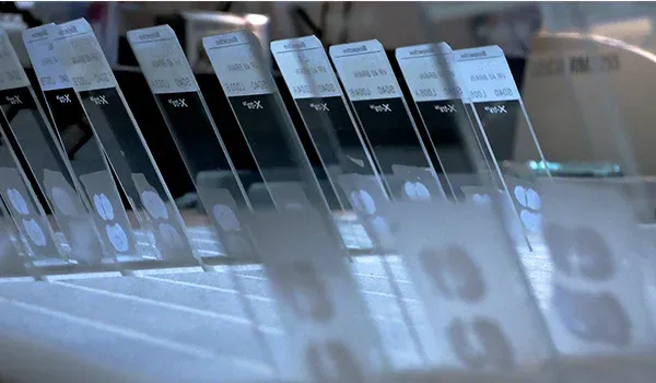
We work with both frozen and FFPE tissue. We provide a comprehensive range of services, including tissue processing, paraffin embedding, and tissue sectioning.

A close-up, angled view of microscope slides, each with a specimen spot and a label

Immunohistochemistry (IHC)

We perform high-quality IHC studies using a range of markers tailored to our CNS disease models.

Immunohistochemistry (IHC) staining of the mouse substantia nigra

Multiplex Immunofluorescence (IF)

We combine multiple immunofluorescence markers on a single tissue section.

Multiplex IHC image

Slide Scanning

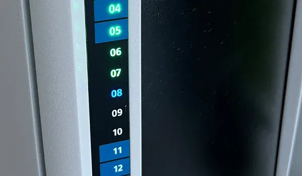
We use automated, high-throughput slide scanners to digitize entire tissue sections.

Slide Scanning Equipment used for Histology & Tissue Analysis

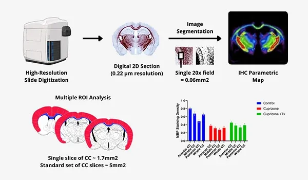
Quantitative Digital Image Analysis

We leverage our proprietary PERMITS™ software to derive quantitative measures from digitized tissue sections.
Quantitative Digital Image Analysis Process in preclinical research studies

Spatial Biology

We analyze spatial relationships on multiplex immunofluorescence images using sophisticated algorithms.

A composite of three different microscopic images or scans, each illustrating a different aspect of biological tissue analysis, potentially within the context of neurobiology

More Information

Let us know what you’re interested in. Our team will be happy to discuss with you!

Email us at i[email protected] or simply complete & submit the form below. 

Name*
Email*
Phone Number*
Affiliation (Company/University)*
Message*

Your privacy is important to us. We will protect your data as outlined in our Privacy Notice.

I agree to the terms in the Privacy Notice*

We use necessary cookies to make our site work. We also use other cookies to help us make improvements by measuring how you use the site or for marketing purposes. You have the choice to accept or reject them all. For more detailed information about the cookies we use, see our Privacy Notice.