Preclinical WebsiteClinical Website

Positron Emission Tomography (PET)

Preclinical PET imaging allows for metabolic and molecular imaging in rodent models. PET imaging is currently used in clinical trials of neurodegenerative diseases to assess glucose metabolism, β-amyloid burden, tau pathology, and dopaminergic terminal density.

At Biospective, we have demonstrated regional glucose hypometabolism is several of our mouse models, including our alpha-synuclein Parkinson's disease model and TDP-43 ALS mouse model. We can also work with a range of other radiolabeled tracers for PET studies in mice and rats.

Two small square panels, each depicting what appears to be a Positron Emission Tomography (PET) scan, a type of imaging test that helps reveal how tissues and organs are functioning

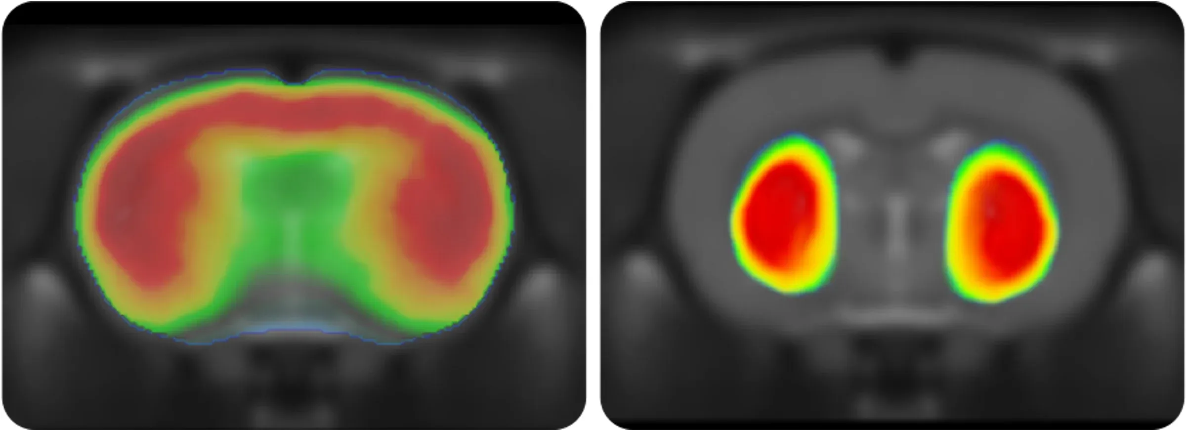
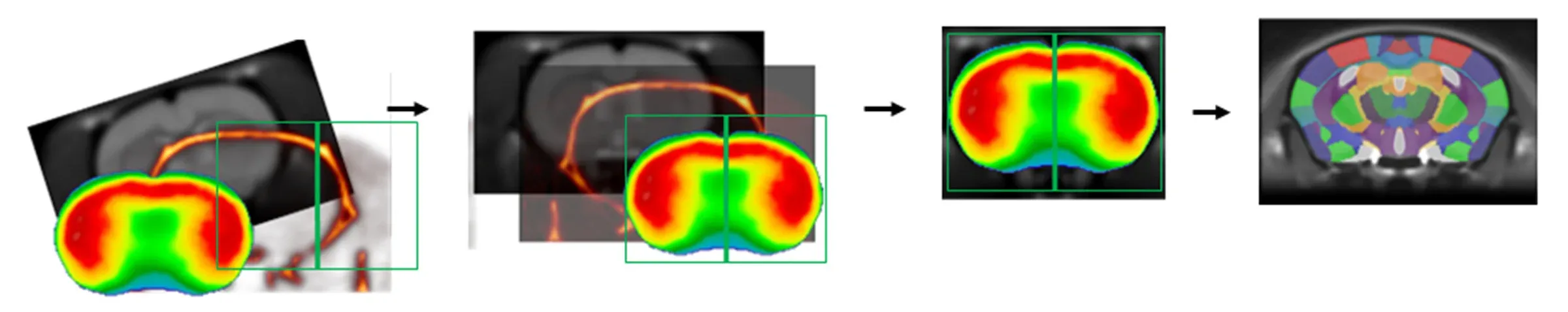
A key to successful PET imaging is rodent model is accurate analysis of measures, such as SUVR, from specific brain regions or via voxelwise analysis. We have developed sophisticated, image registration algorithms, as part of our our proprietary NIGHTWING™ software platform, to align rodent brain PET images with MRI scans to allow for fully-automated analysis.

In Vivo Imaging - PET Processing Pipeline

Learn more about our Positron Emission Tomography (PET) Services.

Discover more of our In Vivo Imaging Services

FAQs

What is SUVR?

SUVR stands for Standardized Uptake Value Ratio. It is a measure of the radiotracer uptake and/or binding in w a specific region normalized to a reference region. In rodent models, typical reference regions for brain PET SUVR analysis include whole brain, cerebellum, and brainstem.


What is voxelwise analysis?

A voxel (or volume element) is a pixel with a third-dimension. In brain imaging, rather than analyzing the data from an entire region-of-interest (ROI), we can perform statistical comparisons on a voxel-by-voxel basis. With this approach, we typically generate statistical parametric maps, such as t-statistic maps. Voxelwise analysis is often an excellent way to explore imaging data without a priori hypotheses.


How is PET different from MRI?

PET and MRI complement each other. While MRI is typically used to glean information about brain structure or tissue biophysical properties, PET can provide information about brain metabolism and molecular neurobiology (e.g. receptor expression).


What is the spatial resolution of preclinical PET scans?

The spatial resolution of our PET scans are <1 mm. While that is lower than the resolution of MRI scans, it is still sufficient to evaluate measures, such as cerebral glucose metabolism, in different brain structures, such as the cerebral cortex, hippocampus, striatum, thalamus, olfactory bulb, cerebellum, and brainstem.


More Information

Let us know what you’re interested in. Our team will be happy to discuss with you!

Email us at i[email protected] or simply complete & submit the form below. 

Name*
Email*
Phone Number*
Affiliation (Company/University)*
Message*

Your privacy is important to us. We will protect your data as outlined in our Privacy Notice.

I agree to the terms in the Privacy Notice*

We use necessary cookies to make our site work. We also use other cookies to help us make improvements by measuring how you use the site or for marketing purposes. You have the choice to accept or reject them all. For more detailed information about the cookies we use, see our Privacy Notice.