Histology & IHC/mIF Services
We have a state-of-the-art histopathology facility specializing in tissues collected from rodent models of neurological diseases.
Neuroscience Specific Markers
We offer a growing portfolio of markers to support research in neurodegeneration and neuroinflammation. Explore our current offerings below:
Tissue Processing
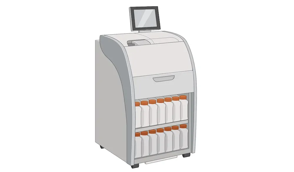
Frozen tissue is typically mounted in OCT blocks following fixation and cryoprotection. For formalin-fixed tissue that will be embedded in paraffin blocks (FFPE tissue), we process the tissue through graded alcohols and xylenes for dehydration, followed by infiltration with paraffin wax. We use an automated tissue processor which handles large numbers of tissue samples and provides consistent processing.
Paraffin Embedding
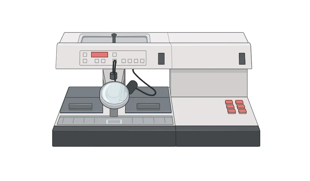
The dehydrated and paraffin-infiltrated tissues provided by our automated tissue processor are then embedded into paraffin blocks. We use a paraffin embedding station to properly orient the tissue in molds, and then fill the molds with paraffin wax with appropriate melting point and hardness to facilitate high quality tissue sectioning and subsequent staining.
Tissue Sectioning
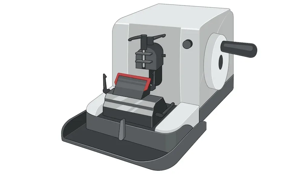
We use digital microtomes for tissue sectioning. These highly accurate instruments allow for sectioning at a wide range of thicknesses. For FFPE tissue, we typically section at 5 µm thickness. We couple our microtomes with cooling/freezing stages. This strategy not only allows us to rapidly cut FFPE tissue, but also provides an excellent set-up for frozen tissue sectioning. We typically section frozen tissue at 20-30 µm thickness.
Slide Scanning
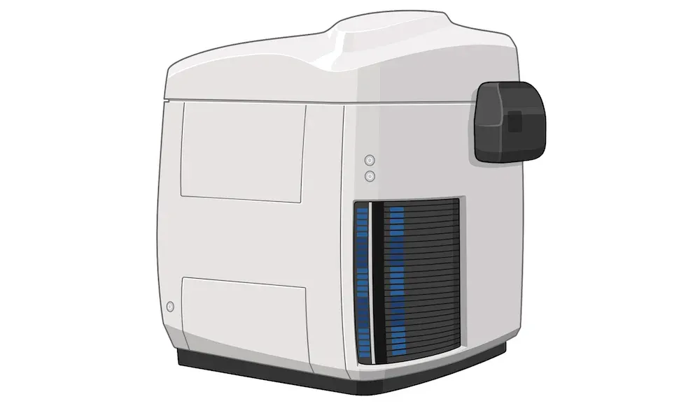
Automated slide scanning for digital microscopy streamlines the process of analyzing tissue samples by employing sophisticated technology to digitize entire microscope slides. Slide scanning can be performed in brightfield or multi-channel immunofluorescence modes.
At Biospective, we have multiple, high-throughput digital slide scanners, including the ZEISS Axioscan 7 system. Slides are scanned at sub-micrometer spatial resolution, typically with 20x or 40x objectives.
These scanners are ideally suited for digitization of multiplex immunofluorescence slides. We use a high-quality LED light source and filters in the range of the fluorophores typically used multiplexing.