Preclinical WebsiteClinical Website
Last Updated Date: July 06, 2024

What constitutes a good research model of ALS for drug development?

The efforts to develop new, effective therapeutics for Amyotrophic Lateral Sclerosis (ALS) (the most common form of Motor Neuron Disease [MND]) have accelerated in recent years. A broad range of targets and mechanisms of action are actively being pursued. Given that in vivo testing in animal models is an essential phase between preclinical in vitro studies and human clinical trials, it is critical that valid models that faithfully replicate multiple aspects of the human disease are used. The optimal animal model of ALS should include several key features described in the infographic below.

Key features of an optimal ALS model

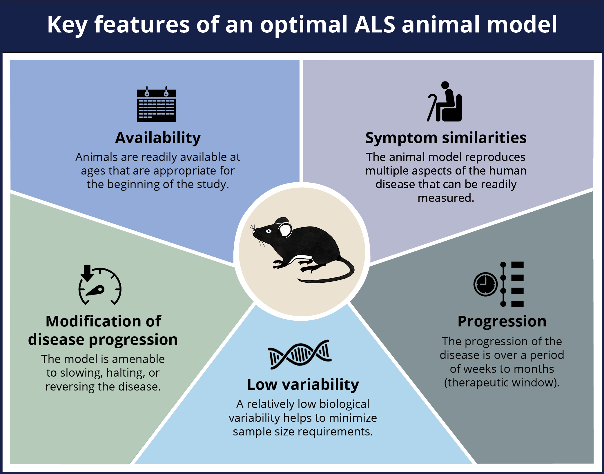
An optimal animal model of ALS should include the following five key features: availability of age-appropriate mice, symptom similarities between the model and the human disease, low variability between the animals, progression of the disease over a period of time, the ability to modify the disease progression in the model.

What are key features of human ALS that should be present in an animal model?

ALS is a progressive neuromuscular disease characterized by loss of upper and lower motor neurons, resulting in muscle weakness and, ultimately, death. The disease can also include a range of non-motor features, including cognitive and behavioral alterations. A pathologic hallmark of both sporadic and familial ALS is TDP-43 proteinopathy, specifically the aggregation of this misfolded protein in the cytoplasm, thereby resulting in toxic loss and gain of function effects. A spatiotemporal pattern of TDP-43 pathology has been proposed by Braak and colleagues and is illustrated below.

Model of the spatiotemporal pattern of spread of TDP-43 pathology in the human brain in ALS (adapted from Braak et al.). 

Like other neurodegenerative diseases, ALS is now viewed as a multi-system disease that is not limited to the central nervous system (CNS), but rather involves many parts of the body. The infographic below highlights some of systems involved in this complex disease.

Multi-system effects of ALS on the human body

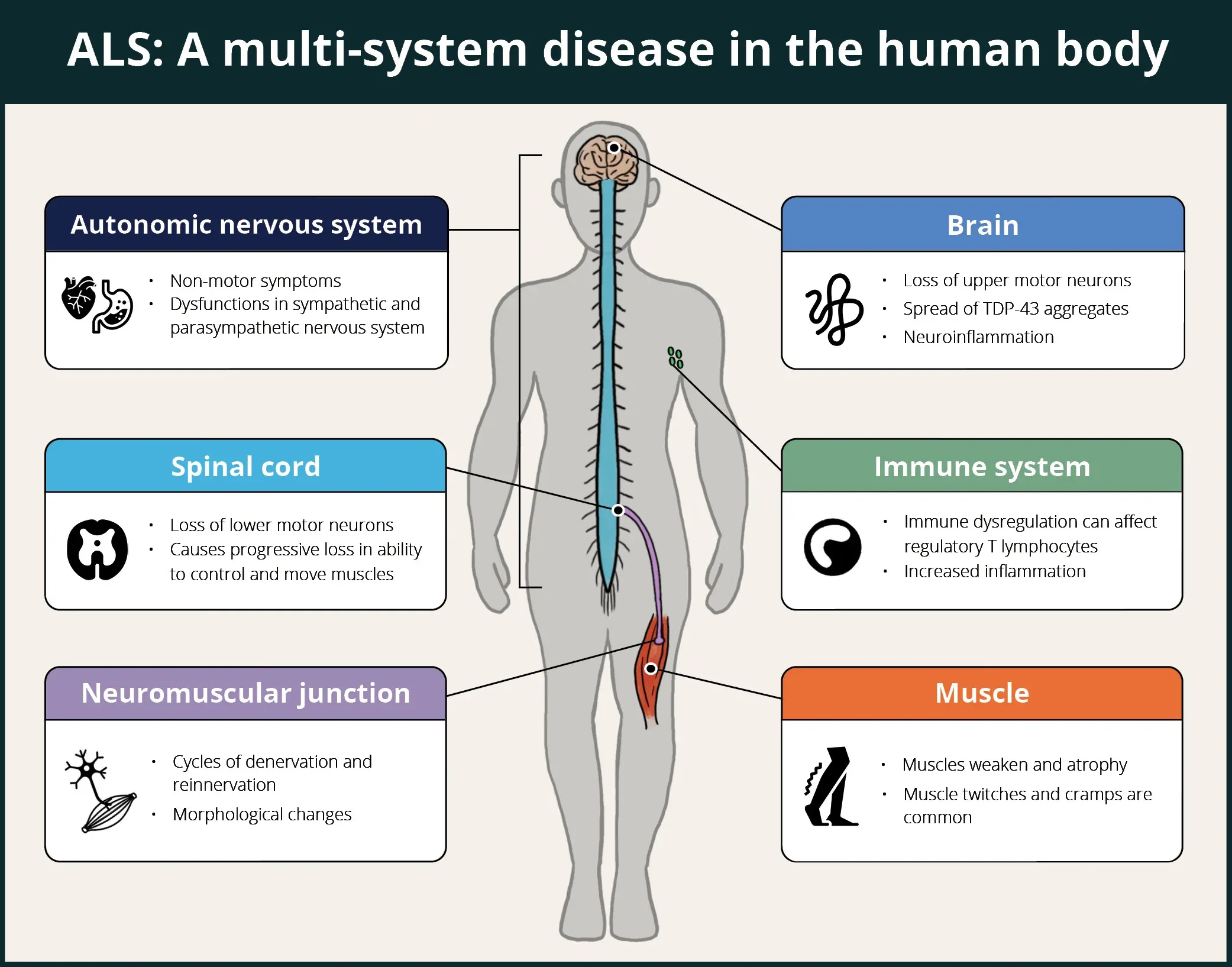
ALS is a multi-system disease in human patients affecting the brain, spinal cord, autonomic nervous system, immune system, neuromuscular junctions, and muscles.

What measures (including translational biomarkers) can be used to evaluate efficacy in ALS models?

A key element to the successful use of ALS mouse models for evaluating the efficacy of therapeutic agents is the use of robust tests that have the sensitivity required to detect disease modification. A multi-modality approach is ideal to capture how a therapy may act at the systems, cellular, and molecular levels. Additional benefit can be gained by employing "translational biomarkers" that could also be leveraged in human clinical trials.

The illustration below provides an overview of some categories of measures that have been used in efficacy studies involving ALS models. Non-invasive imaging measures, such as MRI-derived brain volumes & cortical thickness and glucose metabolism via [18F]FDG PET, are excellent translational biomarkers given that they can be utilized effectively in preclinical animal models studies and human clinical trials. A range of motor function tests to assess general locomotion (e.g. open field), balance & coordination (e.g. rotarod, beam traversal), gait (e.g. CatWalk), muscle strength (e.g. wire hang, grip strength), hindlimb clasping, tremor, and paralysis can be employed to evaluate modification of the clinical phenotype in mouse models. Muscle electrophysiology biomarkers are also clinically translational, where electromyography (EMG) is a standard assessment in ALS  patients. In mouse models of ALS, Compound Muscle Action Potential (CMAP) and Motor Unit Number Estimation (MUNE) can be readily measured from the mouse hindlimb muscles (e.g. gastrocnemius). Neurofilament light has become a widely used fluid-based biomarker in ALS studies, and can also be used as a measure of axonal degeneration/injury & neurodegeneration in ALS mouse models. Finally, a wide range of techniques, such as immunophenotyping of inflammatory cells via flow cytometry, and multiplex immunofluorescence of protein expression coupled with spatial biology analysis, are utilized to assess cellular & molecular changes resulting from therapeutic intervention.

Measures to evaluate efficacy in preclinical ALS studies

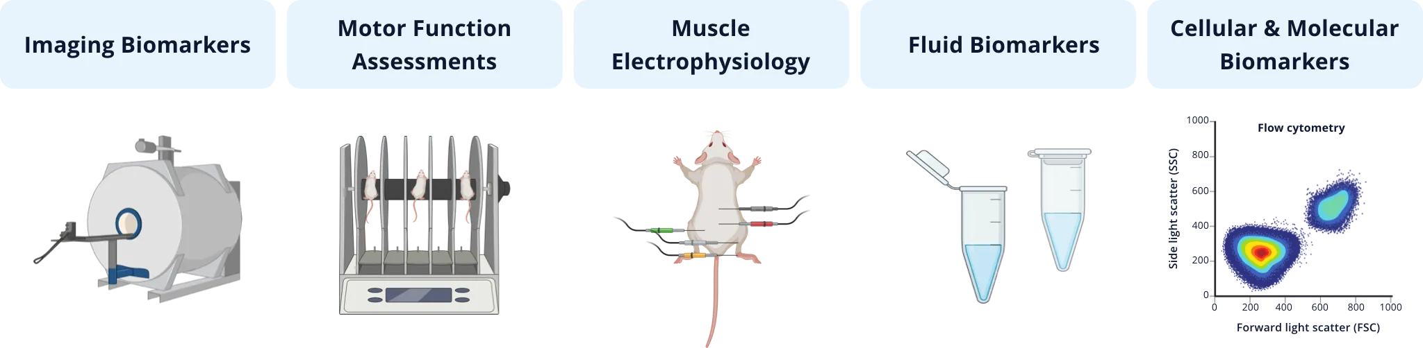
Types of quantitative measures that have been used in ALS preclinical therapeutic efficacy studies involving rodent models.

As an illustrative example of this multi-modality strategy, our team at Biospective has validated a set of these measures in the TDP-43 ΔNLS (rNLS8) transgenic mouse model of ALS, and we are actively evaluating a number of other markers that have been used in human studies. We typically use a combination of these measures, often depending on the target(s) and mechanism(s)-of-action of the therapeutic agent under study to evaluate efficacy. As an example of a translational biomarker, we routinely evaluate brain atrophy in this model using MRI, which shows progressively reduced volume and cortical thickness in the motor cortex, similar to findings in human studies.

Our team would be happy to answer any questions about ALS models or provide specific information about the models that we use for therapeutic efficacy studies.

Discover more about our ALS Models

FAQs

Which in vivo imaging measures have been used in TDP-43 mouse models of ALS?

A number of groups have reported the use of in vivo imaging measures in ALS mouse models. Here is a brief summary of some of the studies, highlighting a number of different clinically translational imaging modalities.

Our group at Biospective routinely performs structural magnetic resonance imaging (MRI) studies to assess regional brain atrophy in the rNLS8 mouse model. We have demonstrated reduced volumes and cortical thickness in the motor cortex. We also use computed tomography (CT) imaging to measure longitudinal muscle atrophy in this hTDP-43 mouse model. We have found significant loss of muscle mass in the gastrocnemius muscle using this non-invasive approach.

Diffusion tensor imaging (DTI) and neurite orientation dispersion and density imaging (NODDI) have been used in the hTDP-43ΔNLS transgenic mouse model, which showed significantly increased intracellular water signal, orientation dispersion, and alteration of DTI metrics. This group also demonstrated impaired glymphatic function in this model using dynamic contrast-enhanced MRI (DCE-MRI), which tracked the clearance of a gadolinium-based MRI contrast agent injected into the cisterna magna.

In this hTDP-43ΔNLS transgenic model, MRI has shown that CSF spaces gradually enlarge over time compared to baseline, while the brain volume was found to decline significantly in comparison to wild-type (WT) mice.  In addition, several brain metabolites were observed by proton magnetic resonance spectroscopy (1H-MRS) to exhibit significant differences between TDP43 and WT mice, including NAA, Cho, and Glx.

In the TDP-43Q331K/Q331K mutant mouse model, structural MRI revealed brain parenchymal volume decrease (with regional volume reductions in cortical, subcortical, and cerebellar regions), as well as an increase in total ventricular volume.

Longitudinal DTI studies have been performed in the TDP-43G298S ALS transgenic mouse model, which revealed disturbed cortical (M1/M2) and callosal microstructure with spared corticospinal tract

In TDP-43A315T transgenic mice, [18F]FDG positron emission tomography (PET) has been used to show significantly lowered glucose metabolism in the motor and somatosensory cortices and elevated metabolism in the region covering the bilateral substantia nigra, reticular, and amygdaloid nucleus between 3 and 7 months of age, as compared to non-transgenic controls. This group also used magnetic resonance spectroscopy (MRS) to explore brain biochemistry, and found significant changes in glutamate + glutamine (Glx) and choline levels in the motor cortex and hindbrain of TDP-43A315T transgenic mice compared to controls.


Which markers can be analyzed from the CSF of TDP-43 ALS mouse models?

Neurofilament light chain (NfL; NF-L) is often analyzed in the cerebrospinal fluid (CSF) from TDP-43 mouse models to assess neurodegeneration and/or axonal injury. It is possible to measure a range of other "biomarkers" (e.g. TDP-43 levels, cytokines, neurotransmitters). The limiting factor is the low volume of CSF that can be collected from mice. At Biospective, we typically obtain ~10 μL of CSF per collection. We have also developed the ability to collect CSF in-life, thereby allowing for multiple collections during the course of the study and facilitating the analysis of multiple disease markers.


How can muscle atrophy be measured in rodent models of ALS? 

There are several methods. The most common approach is to obtain the wet muscle mass. The disadvantage of this method is that it is cross-sectional (postmortem) and can give variable results depending of the dissection of the specific muscle. Care must also be taken for cross-sectional measures where male and female mice are used in the study, as male mice typically have greater muscle mass than females. The inclusion of cross-sectional muscle weights from age-matched male and female mice in a study can have a high biological variability that can obscure a significant drug effect. 

We use computed tomography (CT) imaging coupled with robust automated image segmentation methods. This non-invasive approach allows for longitudinal measures in the same animal and accurate, reproducible measures of different muscles, thereby reducing variability and maximizing the potential to identify a potential therapeutic effect on muscle atrophy.


What is a reasonable sample size for an ALS model efficacy study?

The sample size depends on the biological variability of the model and the methodological variability of the measures. In our studies using the TDP-43 ΔNLS mouse model, we typically use 10-15 mice per group.


References

Benatar, M., Wuu, J., Turner, M.R. Neurofilament light chain in drug development for amyotrophic lateral sclerosis: a critical appraisal. Brain, 146: 2711–2716, 2023; doi: 10.1093/brain/awac394

Braak, H., Brettschnieder, J., Ludolph, A.C., Lee, V.M., Trojanowski, J.Q., Del Tredici, K. Amyotrophic lateral sclerosis — a model of corticofugal axonal spread. Nat. Rev. Neurol., 9: 708-714, 2013; doi: 10.1038/nrneurol.2013.221

Ferrea, S., Junker, F., Korth, M., Gruhn, K., Grehl, T., Schmidt-Wilcke, T. Cortical thinning of motor and non-motor brain regions enables diagnosis of amyotrophic lateral sclerosis and support distinction between upper- and lower-motoneuron phenotypes. Biomedicines, 9: 1195, 2021; doi: 10.3390/biomedicines9091195

Jiang, J., Wang, Y., Deng, M. New developments and opportunities in drugs being trialed for amyotrophic lateral sclerosis from 2020 to 2022. Front. Pharmacol., 13: 1054005, 2022; doi: 10.3389/fphar.2022.1054006

Kraft, S., Mease, C., Jillapalli, D., Fermaglich, L.J., Miller, K.L. Trends in drug development for amyotrophic lateral sclerosis. Nat. Rev. Drug Discov., 23: 99-100, 2024; doi: 10.1038/d41573-023-00199-2

Lin, Z., Kim, E., Ahmed, M., Han, G., Simmons, C., Redhead, Y., Bartlett, J., Emiliano Pena Altamera, L., Callaghan, I., White, M.A., Singh, N., Sawiak, S., Spires-Jones, T., Vernon, A.C., Coleman, M.P., Green, J., Henstridge, C., Davies, J.S., Cash, D., Sreedbaran, J. MRI-guided histology of TDP-43 knock-in mice implicates parvalbumin interneuron loss, impaired neurogenesis and aberrant neurodevelopment in amyotrophic lateral sclerosis-frontotemporal dementia.  Brain Commun., 3: fcab114, 2021; doi: 10.1093/braincomms/fcab114 

Liu, H., Su, S., Longitudinal assessment of TDP43 mouse brain with non-invasive MRI and MRS. FASEB J., 32(supp): 832.5, 2018; doi: 10.1016//10.1096/fasebj.2018.32.1_supplement.832.5

McCombe, P.A., Lee, J.D., Woodruff, T.M., Henderson, R.D. The peripheral immune system and amyotrophic lateral sclerosis. Front. Neuol., 11: 279, 2020; doi: 10.3389/fneur.2020.00279

Müller, H.-P., Brenner, D., Roselli, F., Wiesner, D., Abaei, A., Gorges, M., Danzer, K.M., Ludolph, A.C., Tsao, W., Wong, P.C., Rasche, V., Weishaupt, J.H., Kassubek, J. Longitudinal diffusion tensor magnetic resonance imaging analysis at the cohort level reveals disturbed cortical and callosal microstructure with spared corticospinal tract in the TDP-43G298S ALS mouse model. Transl. Neurodegener., 8: 27, 2019; doi: 10.1186/s40035-019-0163-y

Oprisan, A.L, Popescu, B.O. Dysautonomia in amyotrophic lateral sclerosis. Int. J. Mol. Sci., 24: 14927, 2023; doi: 10.3390/ijms241914927

Weerasekera, A., Crabbé, M., Tomé, S.O., Gsell, W., Sima, D., Casteels, C., Dresselaers, T., Deroose, C., Van Huffel, S., Thal, D.R., Van Damme, P., Hemmelreich, U. Non-invasive characterization of amyotrophic lateral sclerosis in a hTDP-43A315T mouse model: a PET-MR study. Neuroimage Clin., 27: 102327, 2020; doi: 10.1016/j.nicl.2020.102327

Zamani, A., Walker, A.K., Rollo, B., Ayers, K.L, Farah, R, O'Brien, T.J., Wright, D.K. Impaired glymphatic function in early stages of disease in a TDP-43 mouse model of amyotrophic lateral sclerosis. Transl. Neurodegen., 11: 17, 2022; doi: 10.1186/s40035-022-00291-4

Zamani, A., Walker, A.K., Rollo, B., Ayers, K.L., Farah, R., O'Brien, T.J., Wright, D.K. Early and progressive dysfunction revealed by in vivo neurite imaging in the rNLS8 TDP-43 mouse model of ALS. Neuroimage Clin., 34: 103016, 2022; doi: 10.1016/j.nicl.2022.103016


Keywords

Amyotrophic Lateral Sclerosis (ALS): also known as Lou Gehrig's disease, it is the most common form of motor neuron disease and affects the upper and lower motor neurons. This fatal neuromuscular disease is characterized by progressive weakness of the muscles required to move, speak, eat, and breathe.

Brain Atrophy: reduction in volume or thickness of the entire brain or regions of the brain.

Compound Muscle Action Potential (CMAP): summed action potentials of all stimulated motor endplates. 

Motor Unit Number Estimation (MUNE): a technique used to assess the number of functioning motor units within a muscle.

Nuclear Localization Signal (NLS): a short peptide which facilitates the transport of a protein from the cytoplasm into the nucleus of a cell.

Neuromuscular Diseases: disorders that affect the nerves that control voluntary muscles and the nerves that communicate sensory information to the brain.

Neurodegeneration: a complex, multifactorial process resulting in the loss of neurons. 

Neurofilament Light (NfL; NF-L): one of four subunits of neurofilaments, which are proteins found in neurons that provide structure and shape; the neurofilament light level in blood and CSF can serve as marker of neuro-axonal damage.

Spatiotemporal Pattern: a pattern with both spatial and time components.

Transactive response DNA binding protein of 43 kDa (TDP-43):  a highly conserved nuclear RNA/DNA-binding protein encoded by the TARDBP gene involved in the regulation of RNA processing.

Translational Biomarker: a robust indicator of a biological state or process that is measurable in both animal models and humans.


Related Content

Up-to-date information on Amyotrophic Lateral Sclerosis and best practices related to the evaluation of therapeutic agents in ALS animal models.
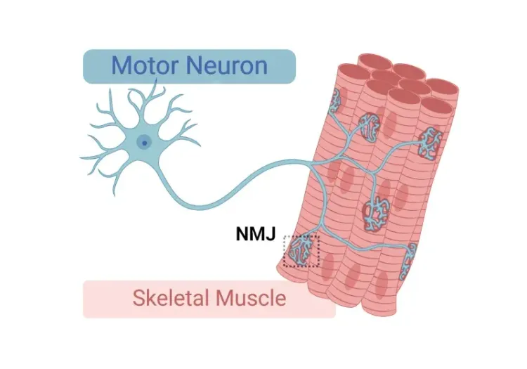
  • TDP-43 ΔNLS (rNLS8) Mice for ALS Drug Development

    This resource provides information about the use of the ΔNLS (deltaNLS, hTDP-43ΔNLS, hTDP-43DeltaNLS, dNLS, TDP43 NLS, rNLS8) TDP-43 transgenic mouse model of ALS for preclinical therapeutic studies.

  • Brain Atrophy Analysis in Rodent Models of Neurodegeneration

    Brain atrophy is a hallmark of many neurodegenerative diseases. MRI is routinely used in clinical trials to non-invasively measure volumetric and cortical thickness changes. We have applied these translational imaging biomarkers to mouse models of ALS and Parkinson’s disease.

More Information

Let us know what you’re interested in. Our team will be happy to discuss with you!

Email us at i[email protected] or simply complete & submit the form below. 

Name*
Email*
Phone Number*
Affiliation (Company/University)*
Message*

Your privacy is important to us. We will protect your data as outlined in our Privacy Notice.

I agree to the terms in the Privacy Notice*

We use necessary cookies to make our site work. We also use other cookies to help us make improvements by measuring how you use the site or for marketing purposes. You have the choice to accept or reject them all. For more detailed information about the cookies we use, see our Privacy Notice.