


Neuroinflammation in the AAV-Tau Mouse Model of PSP
A research study quantifying astrogliosis, microgliosis, and proinflammatory cytokines in Biospective's AAV-hTau (2N4R) mouse model of progressive supranuclear palsy.
 Why Does Neuroinflammation Matter in Progressive Supranuclear Palsy?
- How was Neuroinflammation Induced and Measured in Biospective’s AAV-Tau Model?
- How Much Does Astrogliosis Increase in the AAV-Tau Model?
- How Much Does Microgliosis Increase, and Where?
- Which Cytokines Are Elevated in Midbrain Tissue?
- How Well Do These Readouts Translate to Human PSP?
- How Can These Endpoints Be Used in a Therapeutic Study?
- Summary
Research Study Summary
In brief: We quantified glial reactivity and inflammatory cytokine protein concentrations in Biospective's AAV-Tau mouse model of progressive supranuclear palsy (PSP), addressing an aspect of tauopathy models that is often inferred from tau pathology rather than measured directly. Adeno-associated virus (AAV) vectors expressing human wild-type 2N4R tau were injected into the left substantia nigra pars compacta (SNc) of C57BL/6 mice, AAV-null vector injected into the contralateral substantia nigra; control mice received bilateral AAV-null vector injections, and brains were analyzed 6 weeks post-injection. GFAP and Iba-1 staining densities were significantly increased in the ipsilateral midbrain, SNc, and substantia nigra pars reticulata (SNr), with the largest increases for both markers in the SNc. Neuroinflammatory profiling identified significant elevations in several cytokines including IL-1β, IL-2, and IL-6, while IL-4, IL-10, and KC/GRO were not significantly changed. Together, these findings demonstrate a regionally concentrated glial response accompanied by selective inflammatory cytokine changes. The elevation of IL-1β, IL-2, and IL-6 also aligns with findings reported in PSP patients (Dey, 2026; Rydbirk, 2019) supporting the use of these neuroinflammatory measures as translational endpoints in the AAV-Tau model.
4.3x
Iba-1 microgliosis in the SNc
3.5x
GFAP astrogliosis in the SNc
12x
IL-1β in midbrain tissue
6 of 9
cytokines significantly elevated
4.3x
Iba-1 microgliosis in the SNc
3.5x
GFAP astrogliosis in the SNc
12x
IL-1β in midbrain tissue
6 of 9
cytokines significantly elevated
Fold-change vs. AAV-null controls at 6 weeks post-injection. Jump to the study design ›
The model behind this study
AAV-Tau Mouse Model of PSP & CBD
Human wild-type 2N4R tau delivered to the substantia nigra, producing 4R tau pathology, dopaminergic loss, neuroinflammation, and Parkinsonian motor deficits on a compressed timeline. Our full characterization, interactive microscopy, MRI, and behavioral data can be found on the model page.
Why Does Neuroinflammation Matter in Progressive Supranuclear Palsy?
Neuroinflammation in PSP is not an incidental finding: it is anatomically concentrated in degenerating regions, provides prognostic information beyond clinical rating scales, and is implicated by genetics. Taken together, these findings support incorporating glial and cytokine endpoints into preclinical PSP studies rather than treating them as merely descriptive measures.
PSP is a subcortical-predominant 4R tauopathy
PSP is a sporadic neurodegenerative disease presenting with postural instability and falls, vertical supranuclear gaze palsy, axial rigidity, and frontal-executive impairment (Höglinger, 2017). Neuropathologically, hyperphosphorylated 4-repeat tau accumulates in neurons, in oligodendrocytes as coiled bodies, and in astrocytes as tufted astrocytes. Tau pathology follows a stereotyped anatomical sequence, with early involvement of the subthalamic nucleus, globus pallidus, and substantia nigra (SN), followed by involvement of the striatum, cerebellar dentate nuclei, and cortex (Kovacs, 2020). This distribution has direct implications for model selection: a PSP-relevant rodent model should demonstrate midbrain tau pathology rather than relying solely on findings in the hippocampus or cerebral cortex.
Glial reactivity is associated with regional degeneration
Evidence linking neuroinflammation to PSP pathology comes from both postmortem tissue studies and in vivo imaging. Postmortem image analysis of HLA-DR staining found greater microglial activation in PSP and corticobasal degeneration (CBD) than in control subjects. Microglial burden correlated with tau burden in most regions examined, although the authors cautioned that this relationship was weaker in the brainstem (Ishizawa & Dickson, 2001). TSPO PET studies similarly show increased signal in the basal ganglia, midbrain, and frontal regions (Gerhard, 2006), with elevated thalamic, putaminal, and pallidal signal also reported in PSP-Richardson’s syndrome (PSP-RS; Passamonti, 2018). Postmortem validation indicates that TSPO signal in PSP predominantly reflects microglial rather than astrocytic pathology and correlates with CD68-positive phagocytic microglial burden (Wijesinghe, 2025).
Background on the markers and receptors behind these measurements:
The microglial account is not unanimous
Despite evidence of increased microglial activity in PSP, postmortem findings are not entirely consistent, and other immune responses may also contribute. In FTLD-tau cortex, including PSP, CD16 was the only microglial marker significantly elevated, with the significant subgroup increase occurring in MAPT mutation carriers rather than in PSP (Hartnell, 2024). In the SN, Iba-1 expression did not differ significantly between PSP and controls, whereas CD3+ and CD4+ T-cell densities were markedly higher (Backman, 2025). Midbrain CD8+ T cells have also been described as a distinct feature of PSP (Couto, 2025), and neuroinflammatory profiles may differ by HLA haplotype (Forrest, 2026). Together, these findings suggest that microglial activation is only one component of the inflammatory response in PSP and that its contribution may depend on disease stage, anatomical region, and individual immune characteristics.
Astrocytes participate through two separable processes
Interpreting astrocytic changes in PSP requires distinguishing astrocytic tau pathology from conventional reactive astrogliosis. The distribution of GFAP-positive gliosis does not overlap with that of tufted astrocytes, supporting the distinction between these processes (Togo & Dickson, 2002). At the same time, elevated 1H-MRS myo-inositol and plasma GFAP have been reported in PSP-RS, and reactive astrocytes have been observed in the anterior cingulate cortex despite only mild tau deposition (Hirata, 2024). Consequently, GFAP-based measurements primarily inform the assessment of reactive astrogliosis and should not be interpreted as direct evidence of the tufted-astrocyte tau pathology that characterizes PSP.
Cytokines converge on IL-1β and IL-6
Beyond glial reactivity, human studies provide evidence of inflammatory cytokine dysregulation in PSP, with IL-1β and IL-6 emerging as recurring signals across tissue and biofluid studies. IL-1β transcripts are significantly higher in the SN of patients with PSP than in Alzheimer's disease or control tissue (Fernández-Botrán, 2011), while IL-2 protein is increased in PSP prefrontal cortex (Rydbirk, 2019). Biofluid studies vary by comparator and sample type: CSF TNF-α, IL-1β, and IL-6 are elevated in PSP and MSA relative to Parkinson's disease, but not controls (Starhof, 2018), while elevated plasma IL-1β and IL-6 have been reported relative to controls (Dey, 2026). Cytokine patterns also vary by PSP phenotype and biofluid, with generally lower levels of IL-1β and IL-6 reported in PSP-RS (Madetko-Alster, 2023). Despite this variability, these findings implicate inflammatory pathways that converge on NF-κB and the NLRP3 inflammasome, providing a mechanistic rationale for examining cytokine protein levels alongside glial reactivity in preclinical PSP models.
Cytokine pathway Resources:
Neuroinflammation predicts outcome, though not in one direction
Neuroinflammation may also provide information about disease progression and survival. Baseline brainstem and cerebellar neuroinflammation measured using 11C-PK11195 PET predicted subsequent change on the PSP Rating Scale (PSPRS), whereas MRI volumetry alone did not (Malpetti, 2021). Subcortical TSPO signal and plasma NfL were associated with shorter survival in PSP (Shapiro, 2025), and a peripheral proinflammatory profile was also associated with survival after adjustment for baseline disease severity (Malpetti, 2025). However, these relationships are not uniform: TSPO signal did not correlate with progression across 4-repeat tauopathies (Palleis, 2021), and in β-amyloid-negative CBS, higher global TSPO-PET signal was associated with slower clinical progression (Palleis, 2024). These findings caution against assuming that reducing glial activation will necessarily improve clinical outcomes. Instead, the therapeutic significance of neuroinflammation must be tested using matched readouts.
Genetics points to glia, but not only to microglia
A large PSP genome-wide association study integrating cell-type-specific epigenomic, transcriptomic, and histological data implicated glial activation and the complement system, including the novel risk gene C4A, with prominent oligodendrocyte signal (Farrell, 2024). This broader view of PSP biology is particularly relevant given that anti-tau immunotherapies have demonstrated target engagement without clinical benefit: gosuranemab lowered unbound N-terminal tau in CSF (Dam, 2021), while tilavonemab showed evidence of CSF target engagement but met prespecified futility criteria (Höglinger, 2021). Together, these findings support investigating glial and immune pathways alongside tau pathology, using preclinical models that reproduce relevant anatomical features and allow the resulting glial response to be measured directly.
See our Resource: Microglia, Astrocytes & Tau in Neurodegenerative Diseases
Video 1: The study in 60 seconds. Study design, GFAP and Iba-1 quantification, the nine-analyte midbrain cytokine profile and the patient-to-model readout map in the AAV-Tau mouse model of PSP.
 Click to copy link
How was Neuroinflammation Induced and Measured in Biospective’s AAV-Tau Model?
AAV vectors encoding human wild-type 2N4R tau (AAV-Tau) were injected into the left SNc of young adult C57BL/6 mice by stereotaxic injection using a digital stereotaxic device with an automated microinjector. The contralateral SNc received an AAV-null vector lacking the tau transgene, allowing within-animal comparisons between the tau-expressing and control hemispheres. Control mice received AAV-null vector in place of AAV-Tau. Animals underwent longitudinal behavioral testing and in vivo MRI before being sacrificed 6 weeks post-injection. At study endpoint, astrocytic and microglial reactivity was assessed using multiplex immunofluorescence for GFAP and Iba-1, respectively, followed by quantitative image analysis using Biospective's proprietary PERMITS™ microscopy image analysis platform. Cytokine and chemokine concentrations were measured in ipsilateral and contralateral midbrain homogenates by ultrasensitive multiplex immunoassays.
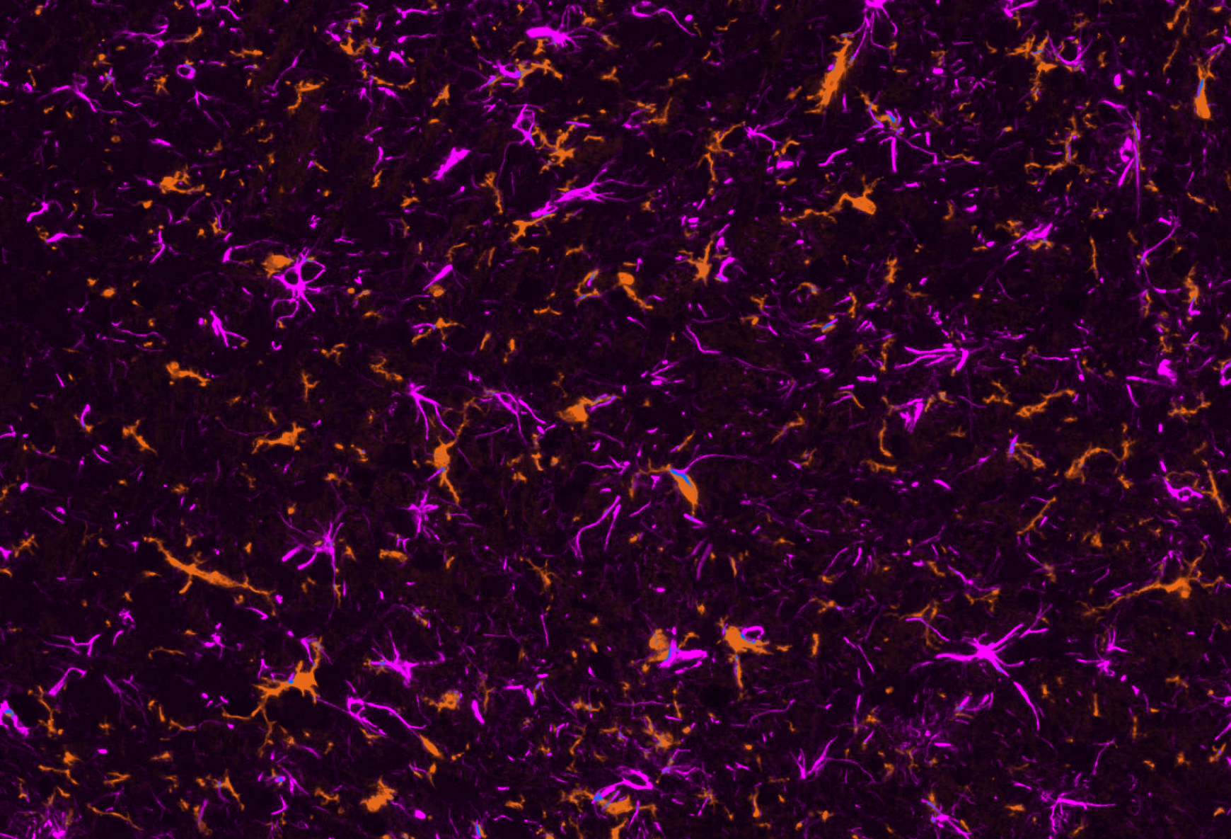
Iba-1 (microglia, orange) and GFAP (astrocytes, violet) double immunofluorescence in the injected SNc of an AAV-Tau mouse. Glial signal is concentrated in the region of highest tau expression.
The phenotype the glial response sits inside
By the 6-week endpoint, AAV-Tau mice exhibited several features of nigrostriatal degeneration alongside the observed inflammatory response. These included phosphorylated and conformationally altered tau in nigral neurons, loss of tyrosine hydroxylase (TH)-positive neurons in the SNc with striatal dopaminergic denervation, significant ipsilateral atrophy of SNc, midbrain and striatum on 7T MRI scans, elevated CSF and plasma NfL, and motor deficits on cylinder, rotarod, tail suspension swing and hindlimb clasping tests. Neuroinflammation was therefore assessed in the context of established nigrostriatal pathology and functional deficits, rather than in isolation. This provides a broader context for interpreting the glial and cytokine measurements.
How Much Does Astrogliosis Increase in the AAV-Tau Model?
GFAP-positive stain density in the ipsilateral hemisphere increased 2.7-fold in the midbrain, 3.5-fold in the SNc, and 1.6-fold in the SNr, relative to AAV-null controls. No significant differences were observed in any contralateral region. This regional gradient indicates that the strongest astrocytic response occurs in the SNc, near the tau injection site, with smaller increases in the surrounding regions.
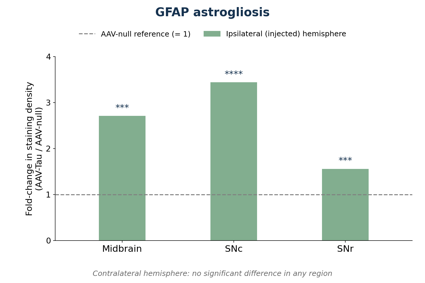
GFAP-positive stain density in AAV-Tau mice expressed as fold-change relative to AAV-null controls in the same region, with the AAV-null value set to 1. Ipsilateral (AAV-Tau- or AAV-null-injected) hemisphere; n = 12 AAV-null and 13 AAV-Tau per region; mean-based fold-change with Welch's t-test; *** p<0.001, **** p<0.0001. Contralateral (AAV-null-injected) values did not differ significantly in any region.
Two aspects of this result are important for interpretation. First, GFAP-positive staining in the contralateral, AAV-null-injected hemisphere of AAV-Tau mice did not differ significantly from that in AAV-null control tissue, despite the same surgery and vector backbone. This finding supports a localized astrocytic response associated with tau expression, rather than a generalized response to surgery or vector exposure. Second, the response varied across anatomical regions, with the largest increase in the SNc and smaller increases in the midbrain and SNr. This pattern is consistent with a spatially concentrated response in regions affected by tau expression, rather than a uniform change throughout the midbrain.
What does this response represent in the context of human PSP? The findings are most consistent with reactive astrogliosis, a process indexed by plasma GFAP and by 1H-MRS myo-inositol. Both measures are elevated in PSP-RS and have been associated with clinical and cognitive scores (Hirata, 2024). Reactive astrogliosis should, however, be distinguished from the tufted-astrocyte tau pathology characteristic of PSP. The latter represents a distinct degenerative process (Togo & Dickson, 2002). Although both processes occur in the PSP brain, GFAP measurements presented here assess reactive astrogliosis rather than the presence of tufted astrocytes. The potential to model tufted astrocytes is currently under investigation by Biospective.
Beyond staining density. Biospective has developed computer vision and machine learning algorithms that classify astrocytes by morphology, distinguishing resting from hypertrophic phenotypes. This approach extends the analysis beyond overall GFAP staining density, helping distinguish changes in astrocyte morphology and state from changes in astrocyte number. See Astrocytes & Amyloid-β Mouse Models of Alzheimer's Disease for an application of this methodology.
How Much Does Microgliosis Increase, and Where?
Iba-1-positive stain density increased 3.4-fold in the ipsilateral midbrain, 4.3-fold in the SNc, and 2.5-fold in the SNr, relative to AAV-null controls. The increase in microglial density exceeded the corresponding astrocytic response in all three regions. The strongest response occurred in the SNc, where tau expression is concentrated. In the contralateral hemisphere, the midbrain showed a nominal 1.3-fold increase, whereas Iba-1 stain density in the contralateral SNc and SNr did not differ significantly from control levels.
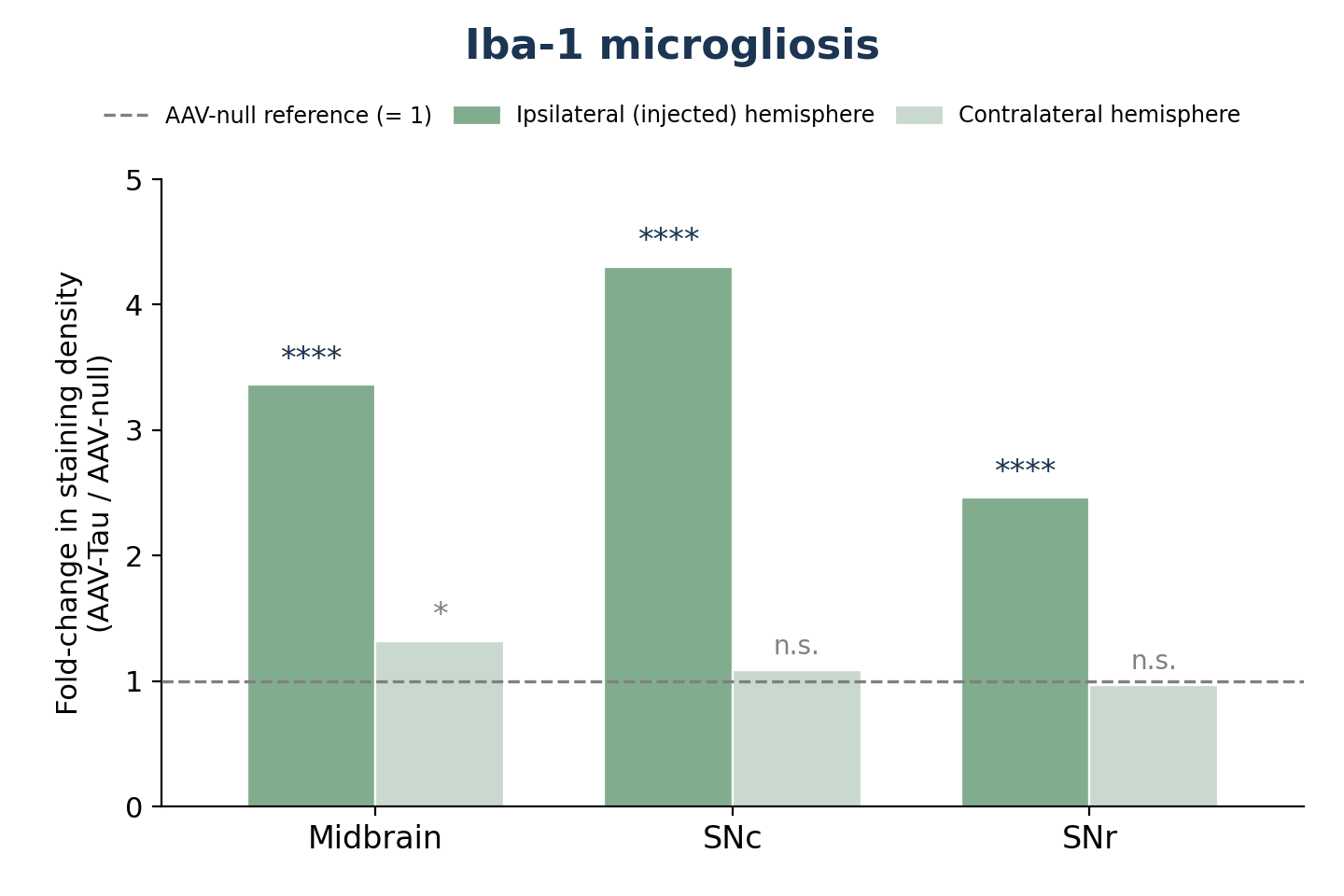
Iba-1-positive stain density in AAV-Tau mice expressed as fold-change relative to AAV-null controls in the same region and hemisphere, with the AAV-null value set to 1. n = 12 AAV-null and 13 AAV-Tau per region; mean-based fold-change with Welch's t-test; * p<0.05, **** p<0.0001; n.s., not significant.
The modest increase in contralateral midbrain Iba-1 staining warrants consideration. This was the only contralateral region showing a significant difference from the AAV-null control group. This signal should be interpreted relative to the AAV-null control group rather than solely against the animal’s own contralateral hemisphere. In contrast, the contralateral SNc and SNr showed no significant differences from control levels.
The regional pattern of microgliosis also aligns with findings from human PSP studies. In this model, microglial increases exceeded astrogliosis and were greatest in the SNc, where tau expression is concentrated. This regional concentration is consistent with human evidence linking microglial burden to regional degeneration (Ishizawa & Dickson, 2001) and TSPO PET studies showing increased signal in the midbrain and basal ganglia (Gerhard, 2006; Passamonti, 2018). Furthermore, postmortem validation has indicated that TSPO signal in PSP predominantly reflects microglial pathology (Wijesinghe, 2025). Therefore, these findings support Iba-1 density as a relevant preclinical readout for investigating microglial responses in PSP.
Density is not state
Although Iba-1 stain density provides a measure of microglial abundance and distribution, it does not fully characterize microglial activation. Iba-1 is a pan-microglial marker, meaning that stain density alone can not distinguish changes in cell number from changes in morphology, process coverage, or reactivity. To address this limitation, Biospective applies a fully automated deep learning approach to classify microglial morphology. This analysis demonstrates a significant shift toward reactive microglial morphology in the SNc of AAV-Tau mice, extending the assessment beyond overall Iba-1 stain density. This distinction is particularly relevant when comparing the model with human PSP findings, where phagocytic CD68-positive microglia, rather than total microglial number, were associated with antemortem PET signal (Wijesinghe, 2025).
See our Resources: Microglia Histology & Analysis & Microglia, Astrocytes & Tau in Neurodegenerative Diseases
Section 4 - Continuation
Need glial state, not just glial density?
We add automated microglia and astrocyte morphology classification, CD68, and other state markers to Iba-1 and GFAP readouts, on your tissue or in a study we perform to evaluate your therapeutic agents in preclinical studies.
Which Cytokines Are Elevated in Midbrain Tissue?
Six of the nine measured cytokines/chemokines were significantly elevated in ipsilateral midbrain homogenates of AAV-Tau mice. IFN-γ increased 24.3-fold, IL-6 increased 14.9-fold, IL-1β increased 12.1-fold, TNF-α increased 4.3-fold, and IL-2 increased 2.2-fold relative to AAV-null controls. IL-5 showed a smaller but statistically significant 1.4-fold increase. In contrast, the anti-inflammatory cytokines IL-4 and IL-10 and the chemokine KC/GRO were unchanged. This profile suggests a selective inflammatory response rather than a uniform elevation across all measured analytes. Histological measurements establish that glial reactivity is increased, while cytokine profiling provides complementary information about the inflammatory mediators present in the tissue.
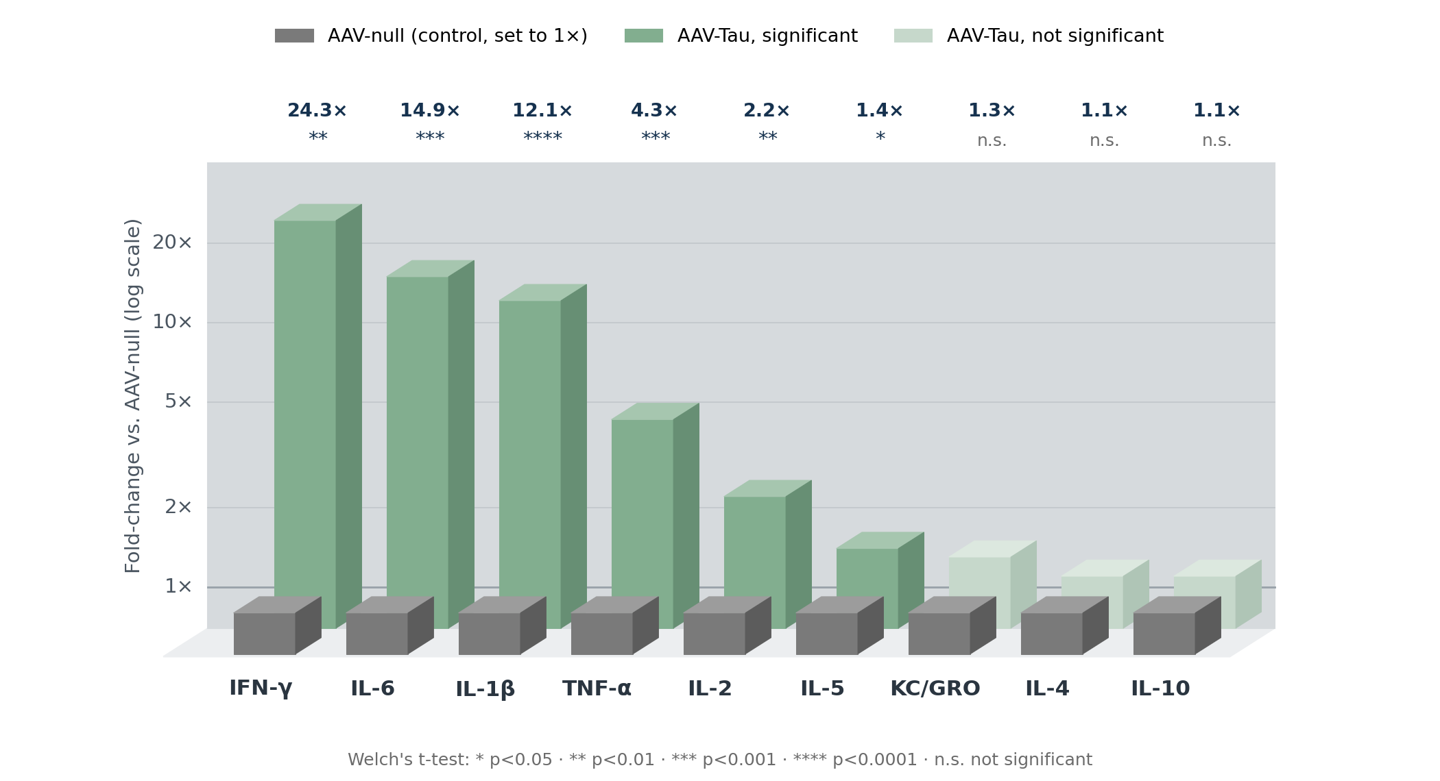
Cytokine and chemokine concentrations in ipsilateral midbrain homogenates of AAV-Tau mice (green, back row) expressed as fold-change relative to AAV-null controls (grey, front row, set to 1×) on a logarithmic axis. Lighter green marks analytes that were not significantly changed. Nine-analyte ultrasensitive multiplex immunoassay, n = 8–10 per group after assay quality control; Welch's t-test; * p<0.05, ** p<0.01, *** p<0.001, **** p<0.0001; n.s., not significant.
Table 1. Cytokine responses in ipsilateral midbrain homogenates of AAV-Tau mice and their reported counterparts in human PSP.
The overall profile is more informative than any single cytokine in isolation. IL-1β and IL-6, two analytes reported across human PSP studies, were also among the most responsive analytes in this model. In contrast, IL-4 and IL-10 did not increase significantly, indicating that the response was not accompanied by a comparable increase in these anti-inflammatory cytokines. IL-2 is the least expected of the significant results because it is not a canonical microglial product. Its elevation in PSP prefrontal cortex has been linked to a shift in peripheral T-cell compartments (Rydbirk, 2019), and IL-2 and IFN-γ together provide cytokine-level points of comparison with the adaptive-immune findings now emerging in human PSP midbrain (Couto, 2025; Backman, 2025). This suggests potential immune-cell involvement but direct evidence is required.
Direct protein quantification across matched tissues and biofluids using ultrasensitive multiplex assays remains uncommon in preclinical tauopathy research, where clinical scores, histology, and gene expression often predominate. Applying the same approach in the EAE mouse model of multiple sclerosis, where cytokine elevation was confined to the CNS with no corresponding plasma signal, illustrates why inflammatory changes should be measured in the relevant compartment rather than assumed.
Related Resources: NLRP3 Inflammasome & Neurodegenerative Diseases & IL-1β & Neurodegenerative Diseases
Section 5 - Continuation
CYTOKINE PROFILING AS A STUDY ENDPOINT
Add multiplex cytokine and chemokine panels to your tauopathy study
The same nine-analyte panel can be applied to brain homogenates, CSF, and plasma from your own cohort, alongside GFAP and Iba-1 quantification in matched tissue.
How Well Do These Readouts Translate to Human PSP?
Each readout in this study has a counterpart measured in PSP patients. The model also enables tissue-level and temporal analyses that are difficult or unavailable in human studies. The section examines how the model aligns with human findings, where it provides additional information, and how it could be extended.
Table 2. Endpoint-level mapping between the AAV-Tau mouse model and measurements made in human PSP.
Where the model adds information that is difficult to obtain in human studies
No protein-level multiplex cytokine data have been published from human PSP substantia nigra: the nigral IL-1β finding is transcript-level (Fernández-Botrán, 2011), the IL-2 protein work is from the prefrontal cortex (Rydbirk, 2019), and the remaining PSP cytokine literature is fluid-based (Starhof, 2018; Dey, 2026). Measurements in ipsilateral midbrain homogenates therefore provide a particularly relevant tissue-level comparison with available human evidence. Separately, human studies of these relationships rely on cross-sectional measurements or a single baseline scan followed by clinical follow-up. By contrast, a model in which tau expression begins at a known time point makes it possible to investigate temporal relationships that autopsy and single-scan study designs cannot directly resolve.
A platform to build on
The current AAV-Tau model captures several features of PSP biology relevant to early therapeutic decisions: 4R tau pathology placed in the substantia nigra, nigrostriatal degeneration, Parkinsonian motor deficits, and a tau-initiated neuroinflammatory response, all on a compressed timeline suitable for preclinical studies. Rather than being a complete replica of human disease, the model provides a foundation for investigating these features. Biospective is exploring ways to extend this unique model to include further PSP-relevant features.
Table 3. Current capabilities of the AAV-Tau model and directions for extending it to capture additional features of human PSP.
Each extension would retain the endpoints described in this section — quantitative glial histology, multiplex cytokine profiling, MRI, fluid biomarkers, and motor testing — so results remain directly comparable with the current dataset. Sponsors with a specific PSP feature in mind can help shape this work; contact us to discuss how a planned extension could support your drug development program.
planning a psp or 4r tauopathy efficacy study?
Talk to the scientists who built and characterized this model
Study design and group sizes powered from the effect sizes on this page
Endpoint selection across glia, cytokines, tau, dopaminergic loss, MRI, and behavior
Ballpark cost and timeline, then a formal proposal
How Can These Endpoints Be Used in a Therapeutic Study?
The combination of a fast, tau-initiated glial phenotype and clinically aligned readouts suits this model to microglia-directed agents, inflammasome and cytokine pathway inhibitors, astrocyte-modulating approaches, and anti-tau candidates whose downstream glial effects are a mechanistic question. Because a robust multimodal phenotype is present by approximately 6 weeks, go/no-go decisions do not require the multi-month timelines of transgenic tauopathy models.
Two built-in comparators
A study can include an AAV-null group that controls for surgery and vector exposure across animals, and every AAV-Tau mouse carries its own AAV-null-injected contralateral hemisphere, which provides a within-animal reference for the same surgery and vector backbone. The AAV-null group is the primary comparator for all endpoints. The contralateral hemisphere adds a within-animal check that the effect is tau-driven and local: it shows no significant GFAP differences from AAV-null controls in every region and for Iba-1 in SNc and SNr, so a treatment effect on the tau-injected hemisphere can be confirmed against both references.
Reading a result in two dimensions
Because glial reactivity and neuroaxonal injury are measured in the same animals, a candidate that suppresses GFAP and Iba-1 without moving NfL, or the reverse, produces an interpretable dissociation rather than an ambiguous one. Given that inflammation carries prognostic information in patients (Malpetti, 2021; Shapiro, 2025), while the direction of that association is not uniform across cohorts (Palleis, 2024), a therapeutic should be judged on whether it moves the glial readouts and the degeneration readouts, not on glial suppression alone.
What Biospective runs end to end
A neuroinflammation study in this model is delivered as a single program, from surgery through to the final quantitative report. Studies can typically be initiated rapidly.
Model induction & in-life
Unilateral AAV delivery by stereotaxic surgery, test article dosing, and longitudinal motor and sensory testing.
Translational in vivo imaging
7T MRI acquisition and automated regional volumetric measures.
Fluid & cell biomarkers
CSF and blood collection feeding multiplex cytokine and chemokine panels and the NfL assay.
Quantitative pathology
IHC and multiplex immunofluorescence (mIF) for microglia and tau, analyzed with PERMITS™.
Summary
Expression of human 2N4R tau in the mouse substantia nigra produces a regionally concentrated glial response accompanied by selective inflammatory cytokine changes, detectable at both the cellular and the protein level within 6 weeks. GFAP and Iba-1 density rise across midbrain, SNc, and SNr, with microgliosis exceeding astrogliosis, and IFN-γ, IL-6, IL-1β, TNF-α, IL-2, and IL-5 rise in midbrain tissue, while IL-4 and IL-10 do not increase significantly.
This profile aligns with IL-1β and IL-6 elevations reported in PSP patients (Dey, 2026) and with the subcortical localization of neuroinflammation on TSPO PET and at autopsy (Ishizawa & Dickson, 2001; Passamonti, 2018), while the adaptive immune response (Couto, 2025) and the tufted astrocyte (Togo & Dickson, 2002) remain outside the model's current scope. With these limitations acknowledged, the model offers a tractable system for investigating whether a therapeutic candidate changes tau-initiated glial biology, using endpoints that map onto the biomarkers used in PSP clinical trials.
FAQs
FAQs
Have a question about this model that isn't answered here?
Our scientific team answers technical questions directly.