


Hypothalamic Atrophy in an AAV-Tau Mouse Model of PSP: Translational Relevance to Human Disease
Hypothalamic volume loss in Biospective's AAV-Tau mouse model, in a structure outside the nigral injection target, mirrors MRI findings in patients with progressive supranuclear palsy (PSP).
 Is the Hypothalamus Atrophied in Progressive Supranuclear Palsy?
- How Does Biospective's AAV-Tau Mouse Model Recapitulate PSP?
- Does the AAV-Tau Mouse Model Show Hypothalamic Atrophy?
- Is Tau Pathology Present in the Hypothalamus of AAV-Tau Mice?
- How Does Hypothalamic Tau in the AAV-Tau Model Relate to Human PSP?
- How Can Hypothalamic Atrophy Be Used in Preclinical PSP Drug Studies?
Research Study Summary
Hypothalamic volume loss is an emerging MRI finding in progressive supranuclear palsy (PSP) that has not been observed in Parkinson's disease (Kassubek, 2026). We assessed whether our image analysis detected this finding in human PSP and whether Biospective's AAV-Tau mouse model can reproduce it.
In baseline T1-weighted MRI scans from the 4RTNI (PSP) and FTLDNI (control) cohorts, bilateral hypothalamic volume was significantly lower in PSP than in controls. In AAV-Tau mice, unilateral injection of human 2N4R tau into the substantia nigra produced significant ipsilateral hypothalamic atrophy seen with in vivo 7T MRI, MC1-positive conformationally altered tau, AT8-positive phosphorylated tau, and TOC1-positive oligomeric tau in the hypothalamus. Because the hypothalamus lies outside the injection target, these results indicate tau-related pathological and structural changes beyond the injected region and add a translational MRI endpoint measurable in the same structure in mice and patients.
A candidate translational endpoint for therapeutic efficacy. Hypothalamic volume is derived by automated, atlas-based volumetry from the same in vivo anatomical MRI scans used for nigral, midbrain, and striatal atrophy endpoints. This allows hypothalamic volume to be assessed without additional scan time for human clinical trials and can also be measured repeatedly in the same animals during preclinical studies. Because the same structure is quantified in patients with PSP, a treatment effect on hypothalamic atrophy in AAV-Tau mice can be expressed as an imaging endpoint with a direct clinical MRI counterpart. This makes hypothalamic volume a candidate efficacy readout for studies of disease-modifying therapies targeting 4R tauopathies.
For full model characterization, see: Tau Mouse Model of PSP & CBD
Video 1. Hypothalamic atrophy in human PSP and in Biospective's AAV-Tau mouse model in 60 seconds: human MRI volumetry, the AAV-Tau study design, mouse MRI volumetry, hypothalamic tau pathology, and multi-domain disease readouts. A full transcript is provided.
Is the Hypothalamus Atrophied in Progressive Supranuclear Palsy?
Biospective analysis of 4RTNI and FTLDNI MRI
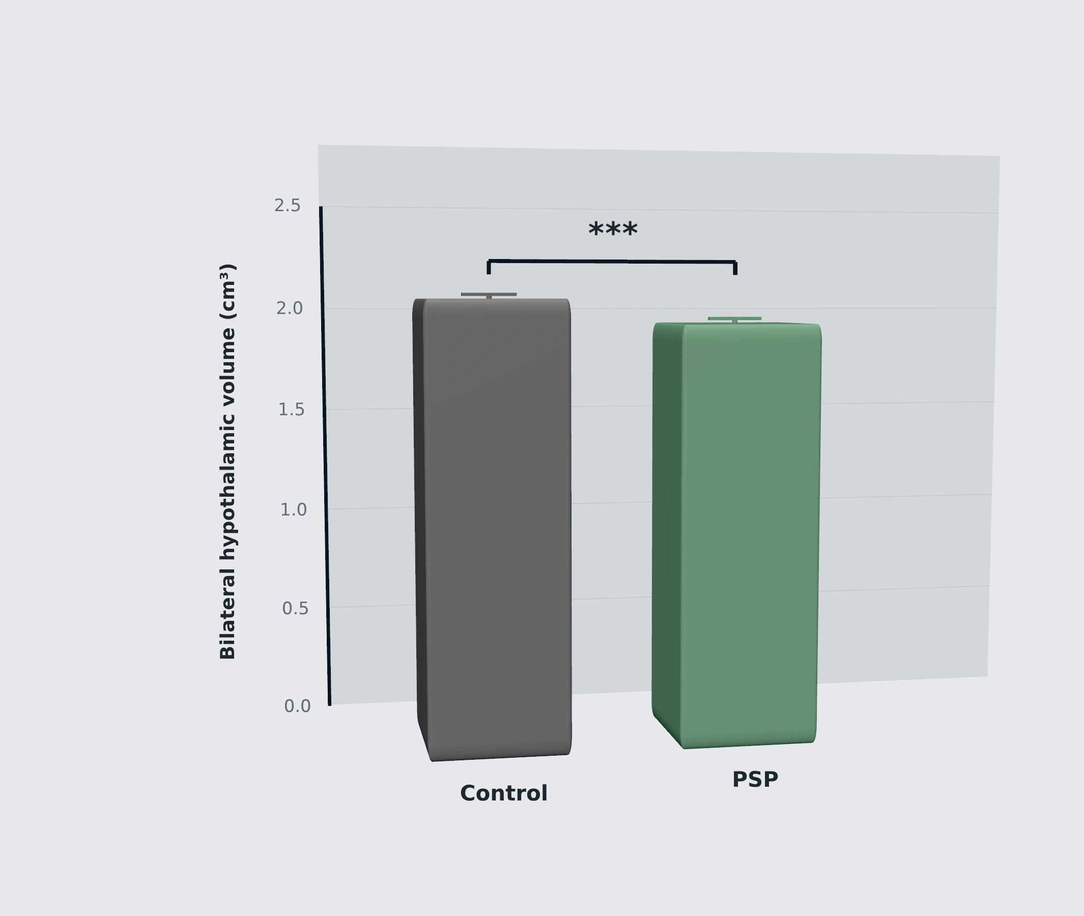
Biospective analyzed baseline 3D T1-weighted MRI scans from participants with PSP in the 4-Repeat Tauopathy Neuroimaging Initiative (4RTNI) and healthy controls from the Frontotemporal Lobar Degeneration Neuroimaging Initiative (FTLDNI) using our fully automated PIANO™ pipeline. The approach is described in our presentation on MRI measures of disease progression for PSP clinical trials. The analysis revealed significantly lower bilateral hypothalamic volume in 51 PSP participants compared to 72 healthy controls (Student's t-test, t(121) = 3.97, p = 0.0001), with a moderate-to-large effect size (Cohen's d = 0.73; Figure 1), consistent with previously published findings using automated segmentation (Kassubek, 2026).
.png)
Figure 1. Bilateral hypothalamic volume (cm³) in healthy controls (gray; n = 72, FTLDNI) and participants with PSP (green; n = 51, 4RTNI) at baseline. Bars show mean ± SEM; Student's t-test, t(121) = 3.97, p = 0.0001, Cohen's d = 0.73; *** p < 0.001. Absolute volumes reflect Biospective's atlas-based definition of the hypothalamus. Biospective in-house analysis of 4RTNI and FTLDNI data.
Data used in this analysis were obtained from the 4RTNI and FTLDNI databases. The 4RTNI and FTLDNI investigators contributed to the design and implementation of these studies and/or provided data, but did not participate in this analysis or in writing this resource.
Consistent with published MRI cohorts
Two independent cohorts analyzed using convolutional neural network (CNN)-based segmentation also showed a smaller hypothalamus in PSP. In a multi-site 3T cohort comprising 78 patients and 63 controls, mean hypothalamic volume was 774 mm³ in PSP versus 817 mm³ in controls. A single-site 1.5T cohort confirmed the reduction while finding no significant hypothalamic volume loss in Parkinson's disease (Kassubek, 2026). The same method detected hypothalamic atrophy in primary lateral sclerosis and ALS (Kassubek, 2025), whereas an earlier MRI volumetry study of 232 Parkinson's disease patients found no significant hypothalamic volume loss (Gorges, 2019). Together, these findings suggest that hypothalamic atrophy may represent an imaging feature that differentiates PSP from Parkinson's disease.
Why this finding has been easy to miss
Many PSP MRI studies report ventral diencephalon measurements rather than the hypothalamus itself. This anatomical label also includes the subthalamic nucleus, substantia nigra, and red nucleus. These regions show early changes in lifespan brain-chart staging of PSP (Planche, 2024) and early atrophy in the subcortical PSP subtype, as identified by machine learning (Scotton, 2023). Consequently, ventral diencephalon volume loss should not be interpreted as direct evidence of hypothalamic atrophy.
The hypothalamus is also absent from the six-stage neuropathological staging system for PSP (Kovacs, 2020) and from the Rainwater Charitable Foundation criteria for neuropathological diagnosis (Roemer, 2022). Its involvement has been underexamined rather than ruled out, underscoring the value of direct measurement of hypothalamic volume.
For more on PSP neuroimaging, see: Imaging Biomarkers for Progressive Supranuclear Palsy & MRI Measures of Disease Progression for PSP Clinical Trials
How Does Biospective's AAV-Tau Mouse Model Recapitulate PSP?
Biospective's AAV-Tau model reproduces several core features of 4R tauopathy with parkinsonian features by delivering human wild-type 2N4R tau into the substantia nigra pars compacta (SNc) of C57BL/6 mice.
Following unilateral stereotaxic injection, mice develop phosphorylated, conformationally altered, oligomeric, and cleaved tau species in the ipsilateral midbrain; loss of tyrosine hydroxylase-positive nigral neurons and striatal dopamine; microgliosis, astrogliosis, and elevated midbrain cytokines; increased plasma tau, GFAP, and NfL; asymmetric motor deficits; and MRI-detected atrophy of the substantia nigra, midbrain, and striatum. In vivo MRI is acquired at week 5, with terminal endpoints assessed at week 6 after injection. Learn more on our AAV-Tau model page.
Section 3 - AAV-Tau study at a glance
AAV-Tau study at a glance
Week 0
Unilateral AAV-hTau (2N4R) injection into the SNc
Weeks 1-5
Longitudinal motor testing
Week 5
In vivo 7T MRI
Week 6
Terminal fluid and tissue collection
Week 0
Unilateral AAV-hTau (2N4R) injection into the SNc
Weeks 1-5
Longitudinal motor testing
Week 5
In vivo 7T MRI
Week 6
Terminal fluid and tissue collection
What can be included in a therapeutic efficacy study in our AAV-Tau model of PSP:
MRI volumetry: Hypothalamus, substantia nigra, midbrain, and striatum
Tau pathology: hTau, AT8, MC1, pTau-T181, PHF1, TOC1, TauΔD421, and aggregated tau
Dopaminergic integrity: Nigral TH staining density and cell counts, striatal TH, and striatal dopamine
Neuroinflammation: GFAP and Iba-1/CD68 assessed using multiplex immunofluorescence; midbrain TNF-α, IL-6, IL-2, IL-1β, and IFN-γ measured using ultrahigh sensitivity immunoassays
Fluid biomarkers: Plasma and/or CSF total tau, GFAP, NfL, and pTau-T181
Motor function: Cylinder test, tail suspension swing test (TSST), rotarod, and SNAP (Simple Neuroassessment of
Asymmetric impairment)
Timeline and endpoints are based on Biospective’s in-house model characterization. Injection target, dosing schedule, and endpoints can be adapted to individual therapeutic programs.
Section 3 Continuation
Why hypothalamic atrophy has not been examined in tau mice
Our literature review found no published studies that directly measured hypothalamic volume by MRI in a tau mouse model. Transgenic tau models do exhibit hypothalamic pathology: hypothalamic and preoptic neurofibrillary tangles have been reported in JNPL3 mice (Lewis, 2000), while suprachiasmatic tau pathology and a lengthened circadian period have been described in rTg4510 mice (Stevanovic, 2017). Progressive sleep and EEG changes have also been reported in rTg4510 mice (Holton, 2020). However, hypothalamic volume was not directly quantified in these studies.
Explore related tau models: Tauopathies Models Overview, Tau Preformed Fibril (PFF) Seeding Mouse Model & A Guide to Mouse Models of Frontotemporal Dementia (FTD)
About the model
Biospective's Tau Mouse Model of PSP & CBD delivers human wild-type 2N4R tau to the substantia nigra via AAV vector, producing tau pathology, dopaminergic neurodegeneration, neuroinflammation, MRI atrophy, and motor deficits within weeks. The model is available for efficacy, mechanism-of-action, and target-engagement studies.
Does the AAV-Tau Mouse Model Show Hypothalamic Atrophy?
AAV-Tau mice show significant hypothalamic atrophy in the injected hemisphere relative to the contralateral hemisphere on in vivo MRI scans.
Native-space hypothalamic volumes were measured with 7T anatomical MRI and our NIGHTWING™ software, part of our preclinical MRI services. See how we measure brain atrophy in rodent models. Across 15 AAV-Tau mice, mean ipsilateral hypothalamic volume was significantly lower than contralateral hypothalamic volume (paired t-test, t(14) = −4.89, p = 0.0002), with a large within-animal effect (Cohen's d = −1.26; Figure 2).
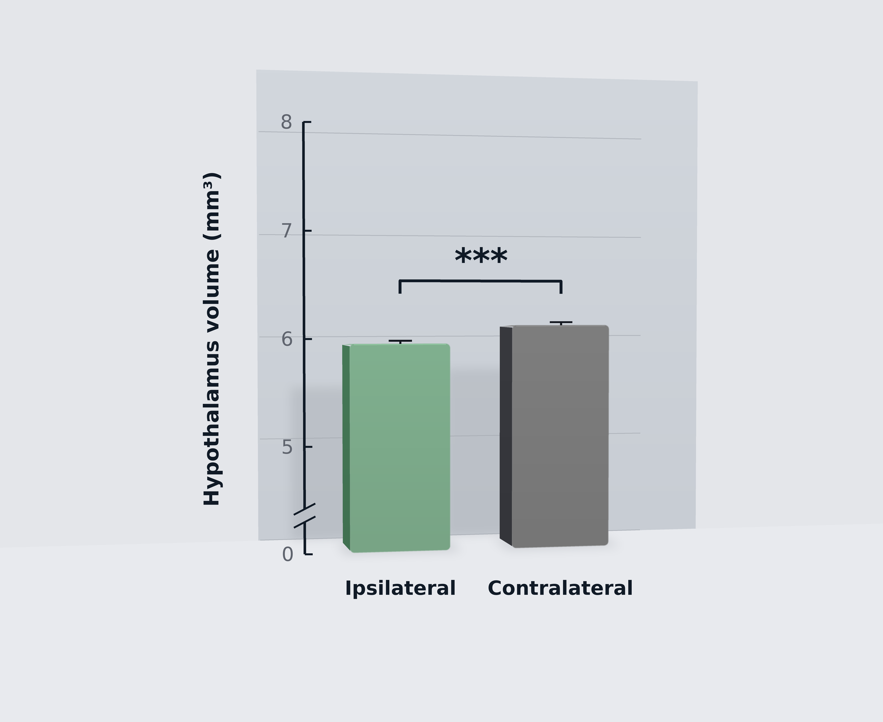
Figure 2. Hypothalamic volume (mm³) in the ipsilateral (injected; left, green) and contralateral (right, grey) hemispheres of AAV-Tau mice, measured using in vivo 7T MRI. Paired t-test, t(14) = −4.89, p = 0.0002, n = 15 mice, with both hemispheres measured in each animal. Cohen's d = −1.26. Bars show mean ± SEM; *** p < 0.001.
The AAV-Tau mice result is consistent with the direction seen in humans. However, the effect sizes are not directly comparable: the mouse effect size reflects a paired, within-animal comparison that accounts for between-animal variation in brain size and registration, whereas the human effect size compares two independent groups. Hypothalamic volume is computed from the same scans already acquired for nigral, midbrain, and striatal atrophy endpoints. Therefore, it can provide an additional structural readout without requiring a separate MRI acquisition when these scans are already part of the study protocol.
Where the hypothalamus fits in the model's MRI profile
On the same in vivo MRI, the ipsilateral substantia nigra, midbrain, and striatum are also atrophied relative to the contralateral hemisphere in AAV-Tau mice. These regions lie within or downstream of the injected nigrostriatal pathway. The hypothalamus provides an additional readout from a region outside the nigral injection target. A single MRI session can therefore capture structural changes within the targeted nigrostriatal system as well as hypothalamic atrophy outside the injection site. For Sponsors, this combination may help assess whether a therapy’s effects extend beyond the targeted nigrostriatal system.
Is Tau Pathology Present in the Hypothalamus of AAV-Tau Mice?
Immunofluorescence reveals multiple pathological tau species in the hypothalamus of AAV-Tau mice, including MC1-positive conformationally altered tau, AT8-positive phosphorylated tau, and TOC1-positive oligomeric tau. MC1 staining is concentrated on the side ipsilateral to the nigral injection.
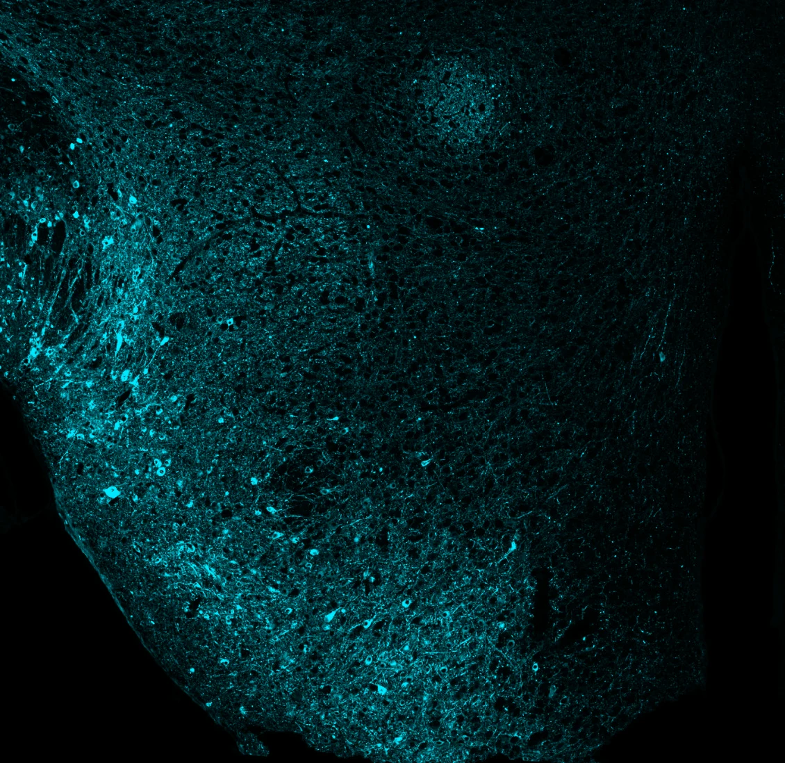
Figure 3. MC1 immunofluorescence (cyan), indicating conformationally altered tau, in a coronal section through the hypothalamus of an AAV-Tau mouse following unilateral injection of AAV-hTau (2N4R) into the left substantia nigra pars compacta. The image shows the ipsilateral hypothalamus, with dense MC1-positive neuronal soma and neurites. The image extends just beyond the midline third ventricle, visible as a dark vertical cleft on the right.
MC1 recognizes a pathological conformation of tau, AT8 recognizes tau phosphorylated at Ser202/Thr205, and TOC1 recognizes oligomeric tau. Detecting all three markers in the hypothalamus indicates that tau pathology in this region includes conformationally altered, phosphorylated, and oligomeric species. In the ipsilateral hypothalamus shown in Figure 3, MC1-positive cell bodies and processes are dense. In the full section, contralateral staining is sparse, consistent with the side of the MRI-detected atrophy. Combining volumetry with tau staining in the same animals allows structural changes to be evaluated alongside tau pathology. These analyses can be quantified using our PERMITS™ microscopy image analysis platform. This provides a way to investigate the relationship between hypothalamic atrophy and tau pathology in the model.
A site of pathology distant from the injection
The hypothalamus lies outside of the injection target. AAV-hTau is delivered to the SNc in the midbrain, yet both the MRI-detected atrophy and the MC1-positive tau are observed in the hypothalamus. These findings indicate that tau-associated pathological changes extend beyond the injected region.
How Does Hypothalamic Tau in the AAV-Tau Model Relate to Human PSP?
Hypothalamic tau pathology in AAV-Tau mice may have translational relevance because human studies have reported tau accumulation and neuronal degeneration in hypothalamic regions involved in sleep and arousal. These changes may contribute to the sleep disturbances associated with PSP (Rastegar-Pouyani, 2026; Oh, 2022; Walsh, 2017).
The same tau isoform class
PSP is a 4-repeat (4R) tauopathy (Kovacs, 2020). The AAV-Tau model expresses human 2N4R tau, placing the expressed tau within the same isoform class that characterizes PSP.
Selective vulnerability of hypothalamic nuclei in PSP
Human post-mortem studies indicate that hypothalamic involvement in PSP varies across neuronal populations. Stereological analysis found 84% fewer neurons in the sleep-promoting intermediate nucleus in advanced PSP than in low-Braak-stage tissue. This nucleus has been proposed as a human counterpart of the rodent ventrolateral preoptic. However, only nine PSP brains were examined (Rastegar-Pouyani, 2026). Tau inclusions were identified in every PSP suprachiasmatic nucleus available for analysis in a sample of five cases (De Pablo-Fernández, 2018). Nevertheless, suprachiasmatic involvement in tauopathies remains understudied (Son, 2024).
Clinical consequences
These pathological findings are consistent with aspects of the sleep and other clinical disturbances observed in PSP. Across Alzheimer's disease and PSP, a greater number of surviving wake-promoting neurons is associated with less total sleep (Oh, 2022). Patients with PSP may sleep fewer than five hours per night (Baschieri, 2026), and polysomnographic findings suggest diminished homeostatic sleep drive (Walsh, 2017). Body weight also declines during the early years of PSP, but not Parkinson's disease (Tsuge, 2017).
Table 1. Hypothalamic features of human PSP and their current status in Biospective’s AAV-Tau model (Biospective in-house data).
Next steps: Biospective's planned hypothalamic characterization
Biospective plans to extend characterization of the hypothalamic phenotype in the AAV-Tau model to determine how closely it reproduces the patterns reported in human PSP. MC1, AT8, and TOC1 staining establishes the presence of pathological tau in the hypothalamus. The next steps are to map the affected nuclei and determine whether their neurons are lost. Planned measures also include non-invasive PiezoSleep sleep–wake recordings.
These studies address open questions in human PSP. Because the hypothalamus is not included as a distinct region in the cited neuropathological staging system (Kovacs, 2020), the timing of hypothalamic involvement in patients remains uncertain. Furthermore, hypothalamic nuclei differ in vulnerability to neurodegenerative pathology; the supraoptic and paraventricular nuclei, for example, are relatively resistant to many neurodegenerative processes (Stewart & Finger, 2021). Mapping nucleus-specific pathology in the model will help determine whether it reproduces the selective vulnerability reported in human studies.
Add a translational hypothalamic MRI endpoint to your next tau study
Biospective conducts fully characterized AAV-Tau efficacy studies in-house, from stereotaxic surgery and dosing to 7T MRI, tau histology, and fluid biomarker analysis, so every readout comes from the same animals.
How Can Hypothalamic Atrophy Be Used in Preclinical PSP Drug Studies?
Hypothalamic volume may serve as a candidate translational MRI endpoint in AAV-Tau efficacy studies because reduced hypothalamic volume has been reported in patients with PSP and has also been observed in this model (Kassubek, 2026; Biospective in-house data).
The same structure, the same modality
Biospective quantifies hypothalamic volume with automated image-analysis pipelines in both rodent and human MRI datasets. For mice, measurements are obtained using 7T preclinical MRI; for human participants, the PIANO™ pipeline is applied to MRI data from the 4RTNI cohort.
Linking structure to pathology and biochemistry
Volumetric measurements can be paired with MC1, AT8, and other tau markers, as well as GFAP and Iba-1/CD68, in hypothalamic sections from the same mice through our tau histology services. In our literature review, we did not identify studies that directly quantified glial pathology in the hypothalamus in human PSP. Quantifying hypothalamic glial markers in the AAV-Tau model could therefore help investigate a potentially understudied aspect of the disease. Reactive glia can be characterized using automated microglial morphology analysis and astrocyte morphology classification.
The same cohort can also provide biochemical readouts measured using ultrasensitive immunoassays and ELISA. In AAV-Tau mice compared with AAV-null controls, terminal plasma total tau, GFAP, and NfL are increased. pTau-T181 is elevated in plasma and ipsilateral midbrain tissue, while ipsilateral midbrain homogenates show raised TNF-α, IL-6, IL-2, IL-1β, and IFN-γ. Striatal dopamine is reduced. These fluid and tissue biomarkers allow sponsors to examine hypothalamic volume alongside systemic markers of neurodegeneration and local measures of tau pathology, inflammation, and dopaminergic integrity within the same animals. Some of these biomarkers also have clinical relevance. Plasma NfL is elevated in PSP and has been associated with clinical progression and brain atrophy (Rojas, 2016). Plasma GFAP can help distinguish PSP from healthy controls and has been associated with brainstem atrophy (Huang, 2023). These findings provide additional points of comparison between preclinical and clinical studies. By contrast, plasma p-tau181 did not distinguish PSP from controls in the cohort reported by Huang et al. (2023).
Functional readouts
Sleep–wake behavior can be assessed non-invasively using PiezoSleep monitoring. This is relevant because sleep disturbances are prominent in PSP (Baschieri, 2026), and sleep measures have been associated with the number of surviving wake-promoting neurons across neurodegenerative diseases (Oh, 2022). Sleep–wake recording and body weight monitoring are planned components of Biospective's ongoing AAV-Tau model characterization and may also be incorporated in Sponsor studies.
Final CTA
Planning a PSP or 4R tauopathy efficacy study?
Our team of scientists can design an AAV-Tau study that combines hypothalamic, nigral, and striatal MRI volumetry with tau pathology, neuroinflammation, fluid biomarkers, and motor testing.
Summary
Hypothalamic atrophy is an emerging MRI finding in PSP, with published imaging cohorts reporting smaller hypothalamic volumes in PSP than in controls and no comparable reduction in Parkinson's disease (Kassubek, 2026). Post-mortem studies have also identified tau pathology and neuronal loss in hypothalamic regions involved in sleep, circadian regulation, and arousal (Rastegar-Pouyani, 2026; De Pablo-Fernández, 2018; Oh, 2019). Biospective's analysis of 4RTNI and FTLDNI MRI data similarly identified reduced hypothalamic volume in PSP. In the AAV-Tau mouse model, significant ipsilateral hypothalamic atrophy was accompanied by MC1-, AT8-, and TOC1-positive tau pathology outside the nigral injection target. Together, these findings support hypothalamic MRI volumetry as a candidate translational endpoint for evaluating therapies in the AAV-Tau model.
We would be happy to discuss how hypothalamic atrophy and our other validated endpoints could support your research program. Please contact us.
FAQs
FAQs