Multiple Sclerosis (MS) Mouse Models
Global preclinical neuroscience CRO specializing in multiple sclerosis models and translational biomarkers.
Validated EAE models & cuprizone mouse models for drug development studies for biotech & pharma.
Biospective's multiple sclerosis animal models (EAE & cuprizone) are optimized for translational neuroscience drug development. These mouse models feature demyelination, inflammatory infiltrates, microgliosis, astrogliosis, and axonal injury. Readouts include clinical & motor assessments, fluid biomarkers (e.g. NfL, cytokines), in vivo imaging (MRI), and quantitative IHC/IF. As a neuroscience CRO, Biospective provides efficacy, MoA, and target-engagement studies in these MS mouse models.
Biospective specializes in mouse models of multiple sclerosis (MS) that replicate key features of human MS, with substantial expertise in neuroimmunology. As a global preclinical contract research organization, we support biotech and pharmaceutical drug development programs using validated multiple sclerosis rodent models for therapeutic efficacy, mechanism-of-action, target engagement, and PK/PD studies across small molecules, antisense oligonucleotides (ASOs), gene therapy (AAVs), antibodies, and other biologics. With fully integrated, end-to-end preclinical services, Biospective facilitates translational MS research from study design through data interpretation.
Multiple Sclerosis Mouse Models – Our Core Expertise
Biospective specializes in disease-relevant MS mouse models for drug development.
Demyelination and inflammation are defining features of multiple sclerosis. As part of our neuroinflammation models portfolio, Biospective has built specialized capabilities around multiple sclerosis animal models.
At Biospective, we use both EAE mice and cuprizone mice to model different aspects of human MS. EAE models autoimmune-mediated demyelination and axonal damage, while cuprizone models demyelination & remyelination without peripheral inflammation. Our in vivo services are focused on reproducibility, well-defined model phenotypes, and the integration of clinical, motor, imaging, biochemical, molecular, and quantitative IHC/IF endpoints to enable comprehensive in vivo MS model efficacy studies and exploration of mechanism-of-action.
EAE Model
Experimental Autoimmune Encephalomyelitis (EAE) is a gold-standard multiple sclerosis mouse model for assessing therapeutic agents targeting autoimmune-mediated CNS disease. The EAE induction is most commonly performed by immunizing mice against myelin-derived antigens, such as MOG, MBP, and PLP. EAE mice model several aspects of human MS.
This model of multiple sclerosis is a primarily T cell-mediated autoimmune disease with several key pathologic features, including demyelination, peripheral inflammation (lymphocytes, macrophages), microgliosis, astrogliosis, axonal injury, and axon degeneration.
Cuprizone Model
The cuprizone MS mouse model of demyelination & remyelination is induced by feeding mice the copper-chelating cuprizone toxin. Cuprizone administration in mice models several aspects of human multiple sclerosis (MS), including demyelination, spontaneous remyelination, oligodendrocyte precursor cell (OPC) proliferation & maturation, astrogliosis, and microgliosis.
The pathology in this demyelination model is primarily limited to the corpus callosum with a highly predictable time course. This demyelination mouse model is well-suited for therapeutic efficacy studies.
Which Features of Biospective's Multiple Sclerosis Disease Models are Translatable to Human Disease?
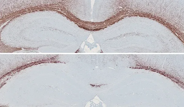
Myelin basic protein (MBP) staining of demyelination in the corpus callosum of the cuprizone mouse model.
Demyelination
MS is the most common demyelinating disease of the central nervous system (CNS), characterized by lesions in both the white matter and gray matter. Both EAE and cuprizone models demonstrate demyelination. EAE causes focal lesions throughout the spinal cord, while cuprizone induces extensive demyelination in specific regions (e.g. corpus callosum).
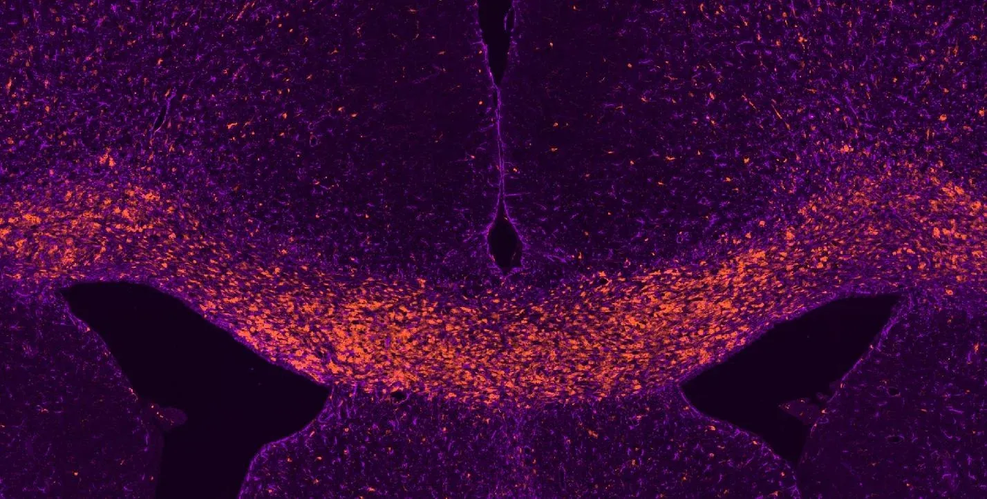
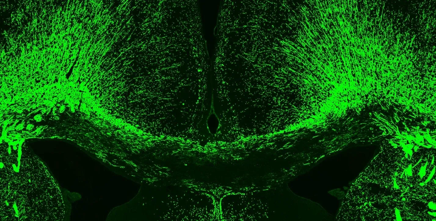
Neuroinflammation (microglia & astrocytes) in the corpus callosum of the cuprizone mouse model.
Activated Microglia & Reactive Astrocytes
Neuroinflammation is a key pathological feature of MS with activated microglia and reactive astrocytes playing key roles in pathogenesis. In our EAE models, microglia & astrocytes are abundant in the spinal cord. In the cuprizone model, neuroinflammation is prominent during the demyelination phase of the disease and resolves as remyelination occurs.
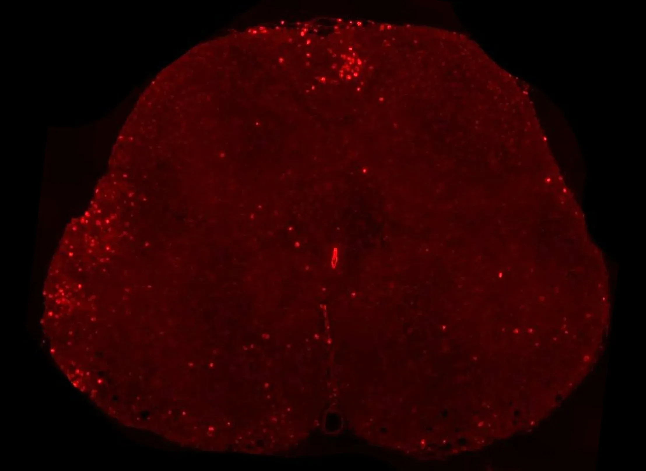
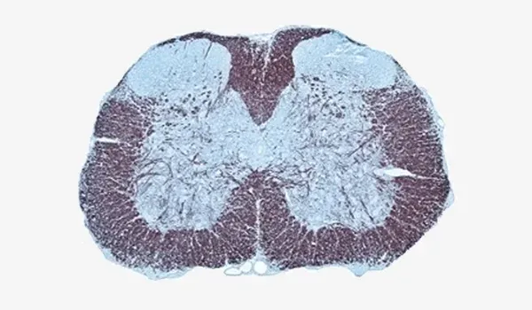
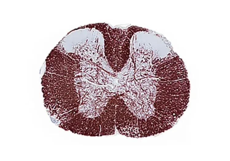
T lymphocytes infiltrating the spinal cord in the MOG35-55 EAE mouse model.
Peripheral Inflammation
EAE is an autoimmune mediated disease. As such, infiltration of T lymphocytes into the spinal cord is a characteristic feature of this model. Further, macrophages are also readily observed in EAE lesions along with resident microglia.
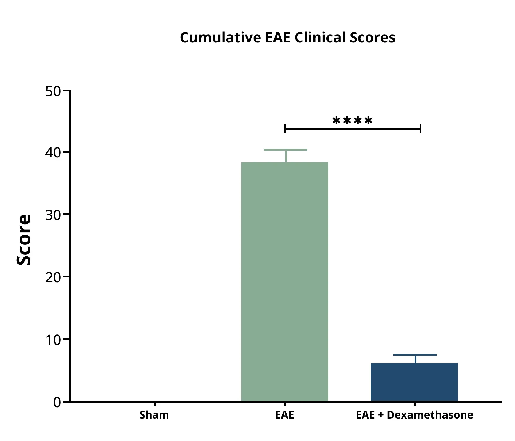
Cumulative EAE clinical scores for sham mice, EAE mice, and EAE mice treated with dexamethasone.
Motor Dysfunction
Progressive motor deficits (e.g. floppy tail, hindlimb paralysis) are a prominent clinical features of the EAE mouse model. A well-defined scoring system (0-5 scale) is typically used to evaluate the extent of motor dysfunction and response to therapeutic intervention.
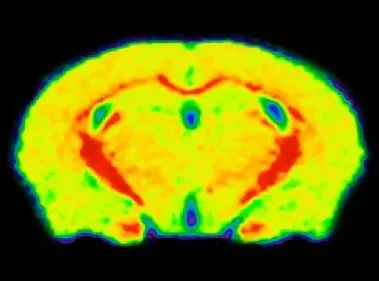
MTR map of a control mouse brain showing high (red) signal in the corpus callosum.
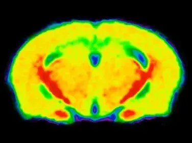
MTR map of a cuprizone mouse brain showing low (green) signal in the corpus callosum.
In vivo MRI Measures
Multi-modality brain imaging biomarkers are widely used in clinical trials of multiple sclerosis. MRI-derived measures of lesion volumes, gadolinium enhancement, lesion biophysical characteristics, and brain atrophy are effective biomarkers of disease progression and response to therapy.
Using whole-brain, high-resolution, anatomical MRI acquisition paired with advanced fully-automated image processing & analysis, we have shown:
- Reproducible magnetization transfer ratio (MTR) measures in the cuprizone model of MS, allowing for longitudinal monitoring of demyelination & remyelination
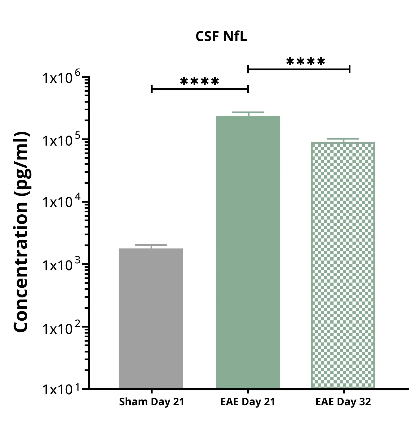
EAE mice show elevated CSF NfL levels at 21 days post-induction compared to control mice.
Elevated Neurofilament Light in CSF & Plasma
Neurofilament light chain (NfL; NF-L) is elevated in the CSF and plasma of MS patients and is routinely used as a fluid biomarker in MS clinical trials (Abdelhak, 2023; Cai, 2018; Konitsioti, 2026). Increased neurofilament light chain levels have also been reported in several preclinical models of MS.
We observe significant increases in plasma and CSF NfL in our MOG35-55 EAE mice. NfL levels serve as a marker of axonal damage & degeneration in this model. See our Resource: Experimental Autoimmune Encephalomyelitis & Axonal Injury.
Why Choose Biospective as Your MS Models CRO?
Biospective is a neuroscience CRO with a focus on MS animal models, strong scientific expertise, and extensive experience conducting preclinical studies with multiple sclerosis models.
-
Specialized Multiple Sclerosis CRO: Focused on MS and neuroinflammatory disease models, not a generalist animal provider.
-
Multiple Validated MS Models: EAE and cuprizone mouse models are readily available for studies.
-
Multiple Sclerosis Expertise: Deep scientific expertise in multiple sclerosis biology and pathology.
-
Integrated Services: Fully integrated preclinical services from study design to data interpretation, ensuring seamless execution.
-
Proven Efficacy Data: Industry-standard EAE & cuprizone models efficacy datasets and extensive historical controls for robust benchmarking.
-
Accelerated Timelines: Rapid study initiation and efficient workflows to compress timelines without sacrificing quality.
-
Translational Biomarkers: Advanced biomarkers (MRI, PET imaging, CSF/blood assays) that bridge preclinical findings to clinical outcomes.
- Flexible & Customized Study Designs: Our scientists work with your team to customize the study design to best fit your goals.
-
Global Support: Experience supporting biotech and pharma clients worldwide, with responsive project management and communication.
Our scientists work as an extension of your internal team, collaborating closely to ensure scientific rigor, reproducibility, and translational relevance at every stage of your MS research program.
End-to-End Multiple Sclerosis Models Preclinical CRO Services
Biospective offers fully integrated preclinical contract research services.
-
Study design & model selection – expert guidance on choosing the right MS model and designing robust studies
-
In vivo efficacy studies – execution of treatment studies with comprehensive monitoring of outcomes
-
Biodistribution & PK/PD – analysis of drug distribution and pharmacokinetics/pharmacodynamics in CNS and periphery
-
Target engagement assays – confirmation that the therapeutic hits its molecular target (e.g. remyelination, reduced inflammation)
-
Clinical & motor scoring – measures such as the EAE Score
-
In vivo multi-modality imaging – MRI, PET, SPECT, fluorescence, and bioluminescence imaging to track disease and treatment effects
-
Immunoassays – biomarker quantification in CSF, blood, and tissue (e.g. NfL, cytokines, chemokines)
-
Immunohistochemistry (IHC) & multiplex immunofluorescence (mIF) – post-mortem tissue staining & quantitative image analysis to assess pathology and therapeutic impact
-
Data analysis & reporting – rigorous quantitative analysis, statistics, and comprehensive reporting by our scientists
This end-to-end approach minimizes handoffs, accelerates timelines, and reduces risk for our sponsors by keeping all aspects of the study with one expert team.
How are MS Mouse Models Used in Drug Development?
We work closely with our biotech and pharma sponsors to:
-
Evaluate therapeutic efficacy and dose-response in MS models
-
Assess target engagement and disease-modifying effects
-
Support translational biomarker strategies, including imaging and fluid biomarkers for clinical readiness
Our MS mouse models are optimized for in vivo testing of multiple therapeutic modalities, including both traditional and advanced approaches:
Small Molecules
-
Brain penetration and PK/PD profile
-
Behavioral efficacy on clinical & motor symptoms
-
Reduction of pathology (demyelination, axonal injury, inflammation)
RNA-Targeted Therapies
- Target knockdown verification (e.g. mRNA or protein level reduction)
-
CNS biodistribution of ASOs/siRNA
-
Translational biomarker readouts to confirm pathway engagement
Gene Therapy & Viral Vectors
-
Transgene expression levels in target regions
-
Regional biodistribution of viral vectors (e.g. AAV spread)
-
Functional rescue or disease modification outcomes (behavioral and pathological improvements)
Antibodies & Biologics
-
CNS exposure and penetration of biologics (e.g. BBB engagement)
-
Aβ and Tau aggregation clearance or reduction
-
Mechanism-of-action validation (target binding, downstream signaling changes)
Contact us to learn more about our characterization of our Multiple Sclerosis mouse models, our validated measures, and our Preclinical Neuroscience CRO services.
Related Content
Up-to-date information on Multiple Sclerosis and best practices related to the evaluation of therapeutic agents in MS animal models.
What is EAE (Experimental Autoimmune Encephalomyelitis)?
An overview of EAE animal models of multiple sclerosis (MS), including pathophysiology and utilization of positive controls for preclinical therapeutic studies.
Experimental Autoimmune Encephalomyelitis (EAE) & Axonal Injury
This resource describes the methods available for measuring axonal damage & axon degeneration, including tissue markers and plasma & CSF neurofilament light chain (NfL; NF-L) levels, in the EAE model of multiple sclerosis (MS).
Demyelination & Remyelination in the Cuprizone Model
An overview of the methods available to measure myelin and oligodendrocytes in the cuprizone demyelination mouse model of multiple sclerosis (MS).
Inflammasome – A Therapeutic Target for Multiple Diseases
An overview of inflammasomes, including their mechanisms of action, roles in diseases, and targeting for drug development.
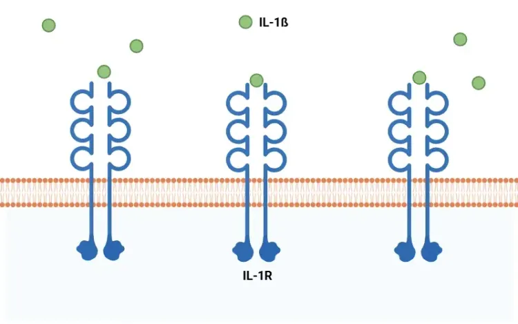
What Is IL-1β (IL-1b)? Function, Signaling, and Biological Role
An overview of IL-1β, including its signaling pathways, involvement in disease mechanisms, and potential therapeutic targets.
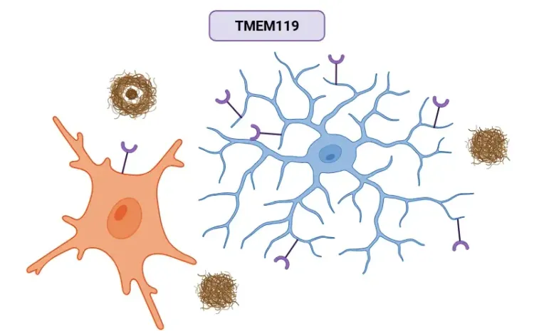
TMEM119 (transmembrane protein 119) and Microglia
An overview of the significance of TMEM119 in labeling microglia and its role in various diseases, including Alzheimer’s disease.
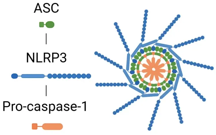
What is NLRP3?
An overview of NLRP3 inflammasome activation triggers, disease associations, and therapeutic targeting strategies.
TNF-α (TNF-alpha) & Microglia in Neurodegenerative Diseases
An overview of the function of tumor necrosis factor-alpha (TNF-α) in microglia and its contribution to the progression of neurodegeneration.
Lysosome Dysfunction in Microglia & Astrocytes
An overview of lysosomal dysfunction in microglia & astrocytes, and its role in neurodegenerative diseases.
TNF-α & (TNF-alpha) Astrocytes in Neurodegenerative Diseases
An overview of TNF-α signaling in astrocytes, its role in neurodegeneration, and therapeutic strategies targeting this pathway..