


Muscle Inflammatory Infiltrates in the "Low Dox" TDP-43ΔNLS Mouse Model of ALS
Muscle Inflammatory Infiltrates in the Low Dox rNLS8 (TDP-43ΔNLS) Mouse Model of ALS
Quantitative multiplex immunofluorescence links CD45⁺ and CD11b⁺ immune infiltrates to neuromuscular junction denervation and CMAP decline in a TDP-43 mouse model of ALS.
 Why does skeletal muscle inflammation matter in ALS?
- How does the Low Dox rNLS8 model reproduce TDP-43-driven neuromuscular pathology?
- Interactive Presentation: muscle inflammation in Low Dox rNLS8 mice
- How were muscle inflammation and NMJ integrity quantified?
- Are CD45⁺ immune cells increased in Low Dox rNLS8 muscle?
- How does NMJ innervation change alongside muscle inflammation?
- Does muscle inflammation track NMJ denervation and CMAP decline?
- Which immune cells make up the muscle infiltrate?
- How does muscle inflammation in Low Dox rNLS8 mice relate to human ALS?
- How can muscle inflammation be used as an endpoint in preclinical ALS efficacy studies?
Research Study Summary
Research Study Summary: Using multiplex immunofluorescence and Biospective’s PERMITS™ image analysis of gastrocnemius muscle from 14 Low Dox rNLS8 (TDP-43ΔNLS) mice and 14 tTA control mice, we found that CD45⁺ immune cell density was roughly twice that of controls, that the infiltrate appears to be predominantly CD11b⁺ myeloid cells, and that across individual Low Dox mice a higher CD45⁺ burden was strongly associated with more fully denervated neuromuscular junctions (r = 0.81) and lower CMAP amplitude (r = −0.71). Muscle inflammation therefore provides a quantitative, mechanistically informative endpoint that complements NMJ innervation and electrophysiology in preclinical ALS efficacy studies.
≈2×
CD45⁺ immune cell density vs tTA controls (p < 0.001)
r = 0.81
CD45⁺ density vs fully denervated NMJs, within Low Dox mice
r = −0.71
CD45⁺ density vs CMAP maximum amplitude, within Low Dox mice
≈2×
CD45⁺ immune cell density vs tTA controls (p < 0.001)
r = 0.81
CD45⁺ density vs fully denervated NMJs, within Low Dox mice
r = −0.71
CD45⁺ density vs CMAP maximum amplitude, within Low Dox mice
Available at Biospective
Low Dox rNLS8 (TDP-43ΔNLS) ALS Mouse Model
In-house breeding colony for rapid start-up and large cohorts
Months-long progression that widens the window to detect disease modification
Validated for muscle inflammation, NMJ innervation, CMAP, CT, grip strength, NfL and MRI
Available at Biospective
Low Dox rNLS8 (TDP-43ΔNLS) ALS Mouse Model
In-house breeding colony for rapid start-up and large cohorts
Months-long progression that widens the window to detect disease modification
Validated for muscle inflammation, NMJ innervation, CMAP, CT, grip strength, NfL and MRI
Study at a glance: muscle inflammation and NMJ integrity in Low Dox rNLS8 mice (Biospective in-house study).
Why does skeletal muscle inflammation matter in ALS?
Skeletal muscle inflammation matters in ALS because immune cells accumulate in patient and rodent muscle around neuromuscular junctions (NMJs), and interventional studies indicate that these cells can accelerate the loss of motor nerve terminals rather than merely clearing up after it (Wang, 2017; Nógrádi, 2025).
Amyotrophic lateral sclerosis (ALS) is defined clinically by the degeneration of upper and lower motor neurons, which produces progressive weakness, paralysis and, ultimately, respiratory failure (Hardiman, 2017). For decades the disease was framed almost entirely around the motor neuron cell body. Spatiotemporal pathology studies changed that view: in SOD1 mutant mice, endplate denervation is already substantial before ventral root axons or spinal motor neurons are lost, and an autopsy from a patient with sporadic ALS showed muscle denervation alongside apparently intact motor neurons (Fischer, 2004). NMJ denervation is also a consistent feature of TDP-43 mouse models, and dying-back mechanisms have been proposed for TDP-43-driven disease, placing the NMJ and its surrounding muscle environment at the centre of disease pathogenesis (Lépine, 2022; Alhindi, 2023).
Muscle is an active participant, not a passive target.
Muscle fibres, fibro-adipogenic progenitors, terminal Schwann cells and resident immune cells all respond to the loss of innervation, and several of these responses feed back on NMJ stability (Shefner, 2023). In denervated muscle, fibro-adipogenic progenitors activate STAT3–IL-6 signalling that promotes atrophy and fibrosis, and blocking this pathway preserved muscle in SOD1 mice (Madaro, 2018). Inflammasome components such as NLRP3 are also altered in ALS mouse muscle and have been explored as a muscle-derived biomarker (Moreno-García, 2021).
Immune infiltrates gather where synapses are lost.
In SOD1G93A rats, myeloid markers including CD11b and CD68 rise in limb muscle as disease advances, accompanied by elevated IL-1β and TNF-α and reactive glial changes adjacent to NMJs (Van Dyke, 2016). In SOD1 mice, complement C5a signalling through C5aR1 on macrophages drives their recruitment into muscle, and deleting C5aR1 reduced both macrophage numbers and endplate denervation while improving grip strength (Wang, 2017). Mast cells and neutrophils also infiltrate paralytic rat muscle and the quadriceps of ALS patients at autopsy, where they associate with motor endplates (Trias, 2017; Trias, 2018).
Most of this evidence came from SOD1 models. The most direct TDP-43-relevant data now come from a study that found leukocyte and macrophage infiltration in muscle biopsies from people with ALS, reproduced the finding in several TDP-43 mouse models from presymptomatic stages onward, and showed that infiltrates concentrate in NMJ-rich muscle regions (Nógrádi, 2025). Proteomic analysis and the enrichment of CCL2-expressing cells around NMJs in hTDP-43 mice and patient muscle implicated the CCL2–CCR2 axis, and local antibody treatment in hTDP-43 mice (with either CCL2-neutralizing antibodies or non-specific IgG) reduced leukocyte infiltration and lessened NMJ denervation (Nógrádi, 2025).
Immune cells are not uniformly harmful, however. Slow-progressing SOD1 mice mount an early muscle response in which infiltrating macrophages adopt a pro-regenerative phenotype that supports myogenesis and preserves CMAP (Margotta, 2023), and in the rNLS8 model microglia are required for full motor recovery once TDP-43 expression is switched off (Spiller, 2018). Measuring both the magnitude and the identity of the immune infiltrate is therefore essential for interpreting what a therapy is doing in muscle.
How does the Low Dox rNLS8 model reproduce TDP-43-driven neuromuscular pathology?
The Low Dox rNLS8 model reproduces TDP-43-driven neuromuscular pathology by expressing human TDP-43 with a defective nuclear localization signal in neurons, which drives cytoplasmic TDP-43 accumulation, NMJ denervation and motor decline (Walker, 2015). Biospective's Low Dox induction paradigm produces this phenotype at a slower, more drug-testable pace than the conventional Off Dox paradigm (Biospective in-house characterization).
The rNLS8 mouse is bigenic: a neurofilament heavy chain (NEFH) promoter-driven tTA line is crossed with a tetO-hTDP-43ΔNLS responder line, so transgene expression in neurons is suppressed by doxycycline and induced when doxycycline is removed (Walker, 2015). In the original description, induction produced insoluble, phosphorylated cytoplasmic TDP-43, loss of endogenous nuclear TDP-43, muscle denervation, motor neuron loss and progressive motor impairment, and returning doxycycline after disease onset allowed muscle reinnervation and functional recovery (Walker, 2015). That reversibility makes the model unusually useful for asking whether a pathway is disease-modifying. Because the transgene is driven by a neuronal promoter, the muscle phenotype described on this page most likely arises downstream of neuronal TDP-43 pathology rather than from transgene expression in muscle itself. For a broader comparison of TDP-43 and other ALS mouse models, see our guide to ALS models for drug discovery and our resource on TDP-43ΔNLS (rNLS8) mice for ALS drug development.
Biospective developed and characterized a reduced-doxycycline (“Low Dox”) induction paradigm that slows progression from weeks to months, widening the window in which a therapy can act. Details of the model, our in-house breeding colony and the full set of validated endpoints are on our rNLS8 TDP-43ΔNLS mouse model page. Previous Biospective data from this model, presented in our NMJ denervation Interactive Presentation, showed loss of SV2A–α-bungarotoxin co-localization, simplified presynaptic architecture and fewer axonal projections, together with reduced grip strength, reduced CMAP amplitude and progressive hindlimb muscle atrophy on longitudinal CT imaging. NMJ denervation has also been documented independently in a Thy1-driven hTDP-43 overexpression model, where it was confined to nerve terminals and tracked motor neuron loss (Alhindi, 2023).
BEYOND THE MUSCLE
See neurodegeneration unfold in the same Low Dox rNLS8 mice
Muscle inflammation is one part of a multi-system phenotype. In Biospective's Low Dox rNLS8 mice, in vivo MRI reveals regional cortical thinning (green and yellow in this animation) alongside cytoplasmic TDP-43, gliosis, rising neurofilament light chain and progressive motor deficits, all measurable within a single study.
Interactive Presentation: muscle inflammation in Low Dox rNLS8 mice
In Low Dox rNLS8 gastrocnemius, CD45⁺ immune cells are distributed throughout the muscle at visibly higher density than in tTA controls, and the Interactive Presentation below lets you inspect this whole-section multiplex immunofluorescence data at full resolution, alongside automated NMJ innervation segmentations and the quantitative results.


 Click to copy link
The presentation is organized so that each microscopy view sits next to the measurement it supports: NMJ structure first, then the CD45⁺ infiltrate in Low Dox and control muscle, the correlation with denervation and CMAP, and finally the CD11b⁺ myeloid component. The sections that follow summarize those results and set them in the context of human disease and drug development.
How were muscle inflammation and NMJ integrity quantified?
Muscle inflammation and NMJ integrity were quantified on fixed-frozen gastrocnemius sections using a five-marker multiplex immunofluorescence panel and Biospective's automated PERMITS™ image analysis, with CD11b staining on consecutive sections and CMAP recorded in the same Low Dox animals.
Animals.
The study compared 14 Low Dox rNLS8 mice with 14 tTA single-transgenic control mice, which carry the NEFH-tTA driver but not the human TDP-43ΔNLS responder transgene (Biospective in-house data).
Multiplex immunofluorescence panel.
Presynaptic nerve terminals: SV2A
Motor axons and terminal branches: βIII-tubulin
Postsynaptic acetylcholine receptors: α-bungarotoxin (α-BTX)
Leukocytes: CD45 (pan-immune-cell marker)
Nuclei: DAPI
Myeloid cells (consecutive section): CD11b
Staining was performed using our automated multiplex immunofluorescence platform and whole sections were digitized on a slide scanner, following the workflow described on our NMJ staining and analysis service page.
Quantitative readouts.
PERMITS™ segmented each channel and derived CD45⁺ cell density (cells/mm² of muscle), the percentage of SV2A signal overlapping α-BTX, presynaptic branching complexity and total axonal projections. Each NMJ was then classified by its SV2A/α-BTX overlap as fully denervated (0–20%), partially innervated (20–80%) or fully innervated (80–100%), and class densities were expressed as endplates/mm² (Biospective in-house classification).
Electrophysiology and statistics.
Maximum compound muscle action potential (CMAP) amplitude was recorded from the gastrocnemius following sciatic nerve stimulation. Group differences were tested with unpaired t-tests and are shown as mean ± SEM; relationships between CD45⁺ density and NMJ or CMAP measures were assessed with Pearson correlation within the Low Dox group, one point per animal (Biospective in-house analysis).
SERVICE
NMJ staining & analysis
Automated innervation classes, presynaptic complexity and axonal projections from whole-section mIF.
SERVICE
Multiplex immunofluorescence
Custom panels such as CD45, CD11b and NMJ markers on a single section, quantified by PERMITS™.
SERVICE
CMAP & MUNE electrophysiology
Clinically aligned motor readouts recorded in the same animals as your histology.
SERVICE
NMJ staining & analysis
Automated innervation classes, presynaptic complexity and axonal projections from whole-section mIF.
SERVICE
Multiplex immunofluorescence
Custom panels such as CD45, CD11b and NMJ markers on a single section, quantified by PERMITS™.
SERVICE
CMAP & MUNE electrophysiology
Clinically aligned motor readouts recorded in the same animals as your histology.
Are CD45⁺ immune cells increased in Low Dox rNLS8 muscle?
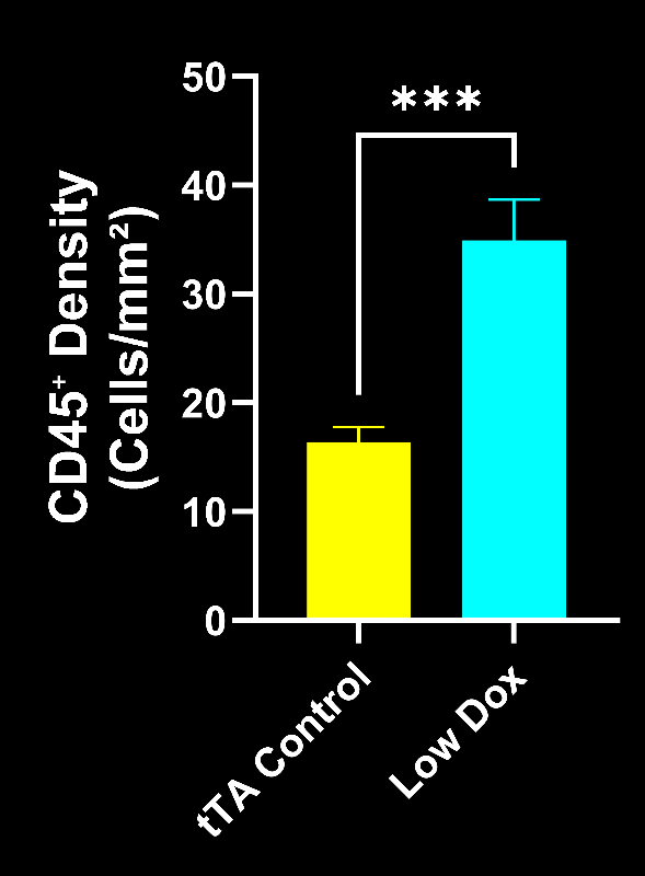
Yes. CD45⁺ immune cell density in the gastrocnemius of Low Dox rNLS8 mice was roughly twice that of tTA controls (p < 0.001; Figure 1), indicating a sustained leukocyte infiltrate rather than baseline immune surveillance (Biospective in-house data).
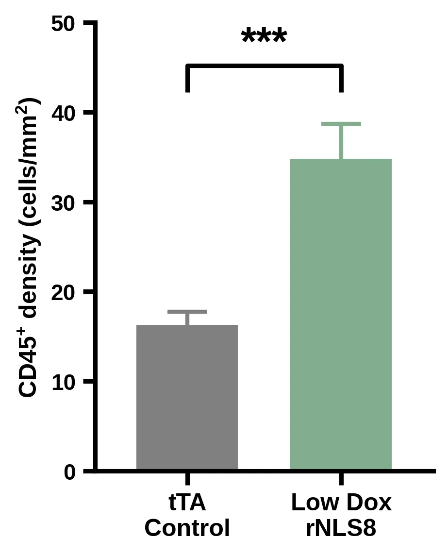
Figure 1. CD45⁺ immune cell density (cells/mm²) in the gastrocnemius muscle of tTA control mice versus Low Dox rNLS8 mice (N=14 per group); mean ± SEM, unpaired t-test, *** p < 0.001. Redrawn in the house palette from Biospective in-house data; group means and SEMs were digitized from the original Prism figures in the Interactive Presentation.
The spatial pattern matters as much as the count. In tTA control muscle, the few CD45⁺ cells present sit mainly in the interstitial spaces between muscle fibres, as expected for resident immune surveillance of healthy skeletal muscle. In Low Dox muscle, CD45⁺ cells are spread throughout the section, which you can confirm by toggling the CD45 channel on the rNLS8 and tTA sections in the Interactive Presentation. Because CD45 labels all leukocytes, this measure captures total inflammatory burden; the identity of the cells is addressed by the CD11b data below (Biospective in-house data). Where the location of immune cells relative to individual endplates matters, our spatial biology analyses can map that neighbourhood directly.
Video Section
WATCH
How we quantify NMJ denervation in ALS mouse muscle
Check out this short narrated overview that walks through Biospective's muscle sectioning, multiplex staining, automated segmentation and innervation classification, using Low Dox rNLS8 and tTA control gastrocnemius as the worked example.
In this video, we provide an overview of our NMJ staining & analysis. It also includes an illustrative example of NMJ denervation in a mouse model of ALS, demonstrating how our services can be used to measure neurodegeneration, monitor disease progression, and evaluate potential therapies in preclinical studies.
How does NMJ innervation change alongside muscle inflammation?
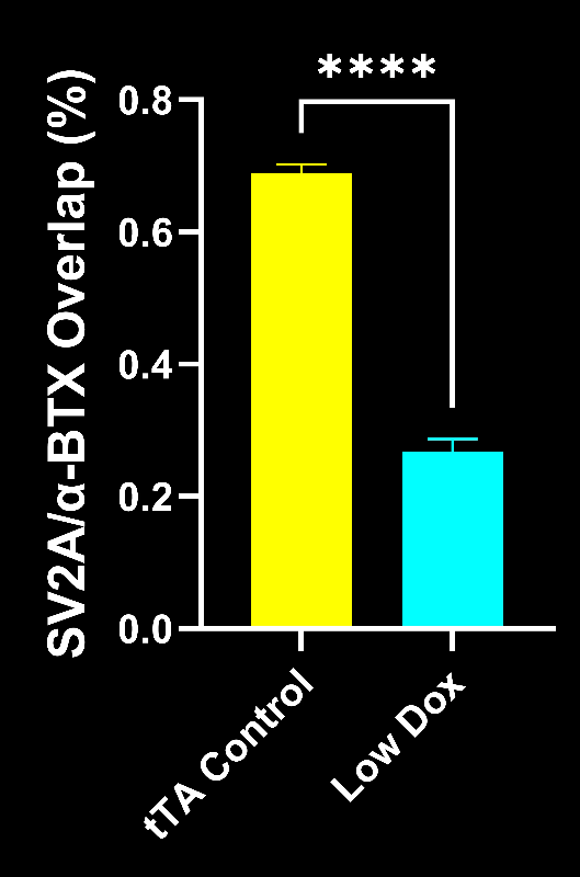
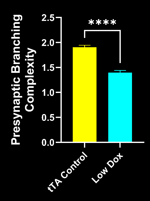
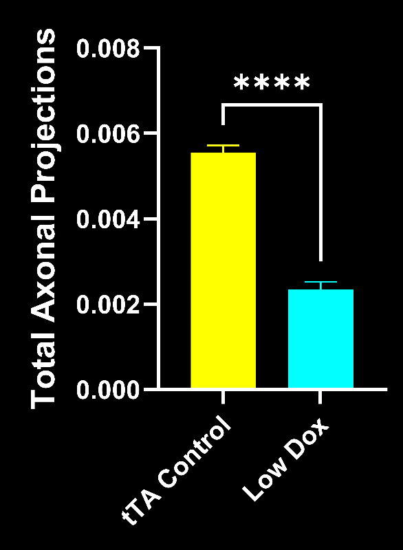
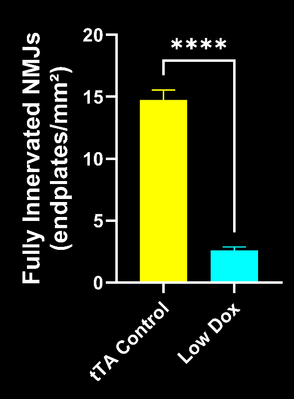
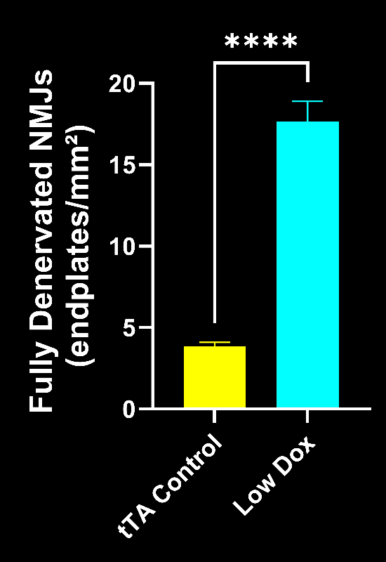
Alongside the inflammatory infiltrate, Low Dox rNLS8 mice showed a pronounced shift from fully innervated to fully denervated NMJs, with reduced presynaptic complexity and fewer axonal projections than tTA controls (all p < 0.0001; Figures 2 and 3; Biospective in-house data).
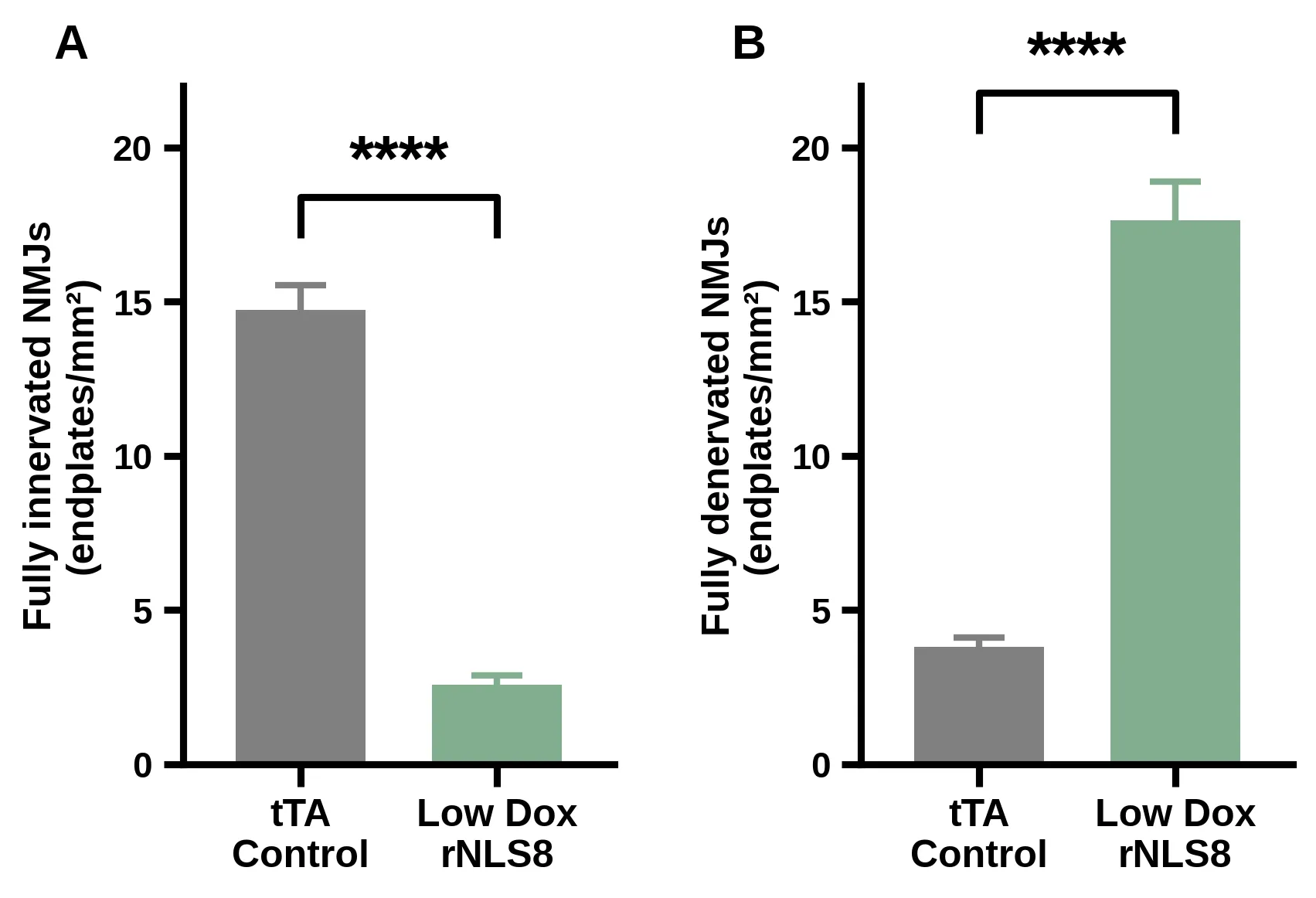
Figure 2. Density (endplates/mm²) of (A) fully innervated NMJs (80–100% SV2A/α-BTX overlap) and (B) fully denervated NMJs (0–20% overlap) in tTA control versus Low Dox rNLS8 gastrocnemius (N=14 per group); mean ± SEM, unpaired t-test, **** p < 0.0001. Redrawn in the house palette from Biospective in-house data (study ID to be confirmed); group means and SEMs were digitized from the original Prism figures in the Interactive Presentation.
Where a program needs tau pathology and frank neurodegeneration alongside amyloid, we run an amyloid-β and tau co-pathology model built on the same ARTE10 background, and the wider Alzheimer's disease model portfolio covers adjacent questions. Our Microglia, Astrocytes & Neurodegenerative Diseases Initiative collects the methodology behind the glial endpoints.
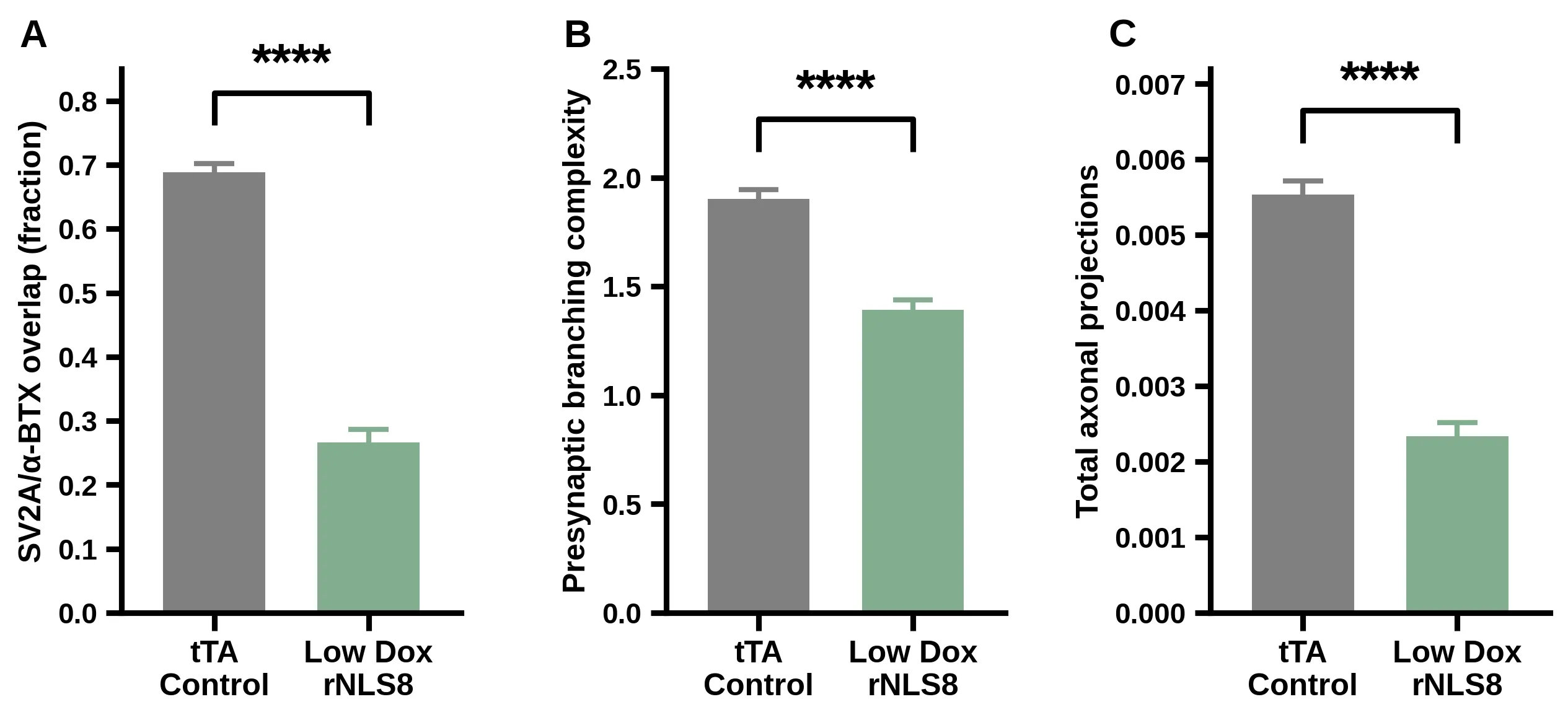
Figure 3. (A) SV2A/α-BTX overlap (expressed as a fraction), (B) presynaptic branching complexity and (C) total axonal projections in tTA control versus Low Dox rNLS8 gastrocnemius (N=14 per group); mean ± SEM, unpaired t-test, **** p < 0.0001 for each. Redrawn in the house palette from Biospective in-house data (study ID to be confirmed); group means and SEMs were digitized from the original Prism figures in the Interactive Presentation.
These NMJ changes reproduce the denervation phenotype described in our earlier NMJ denervation study in this model, and they provide the structural readout against which the inflammatory infiltrate can be compared animal by animal (Biospective in-house data).
Does muscle inflammation track NMJ denervation and CMAP decline?
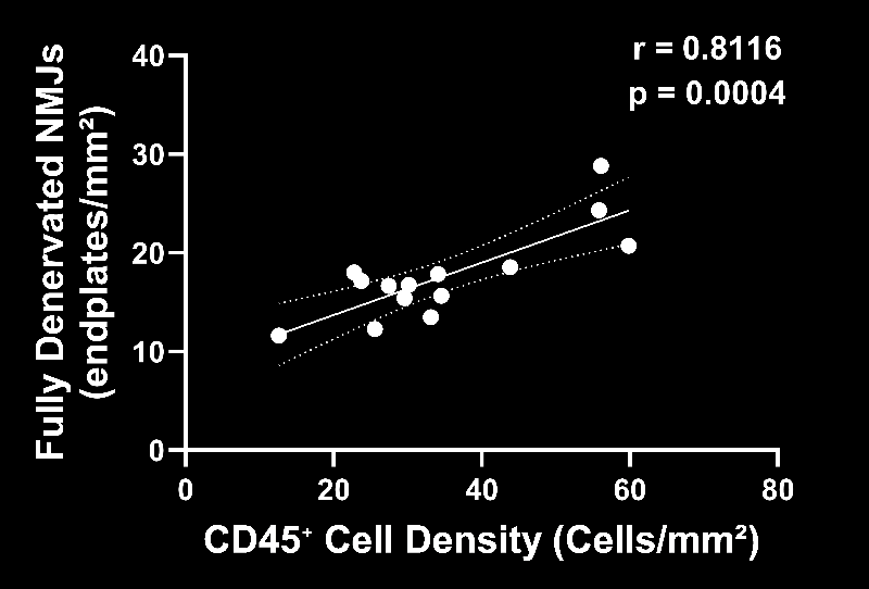
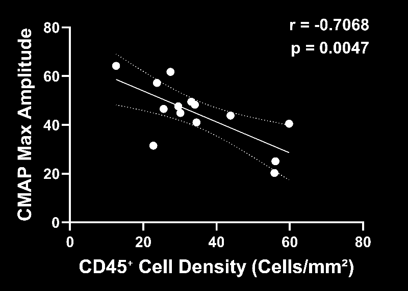
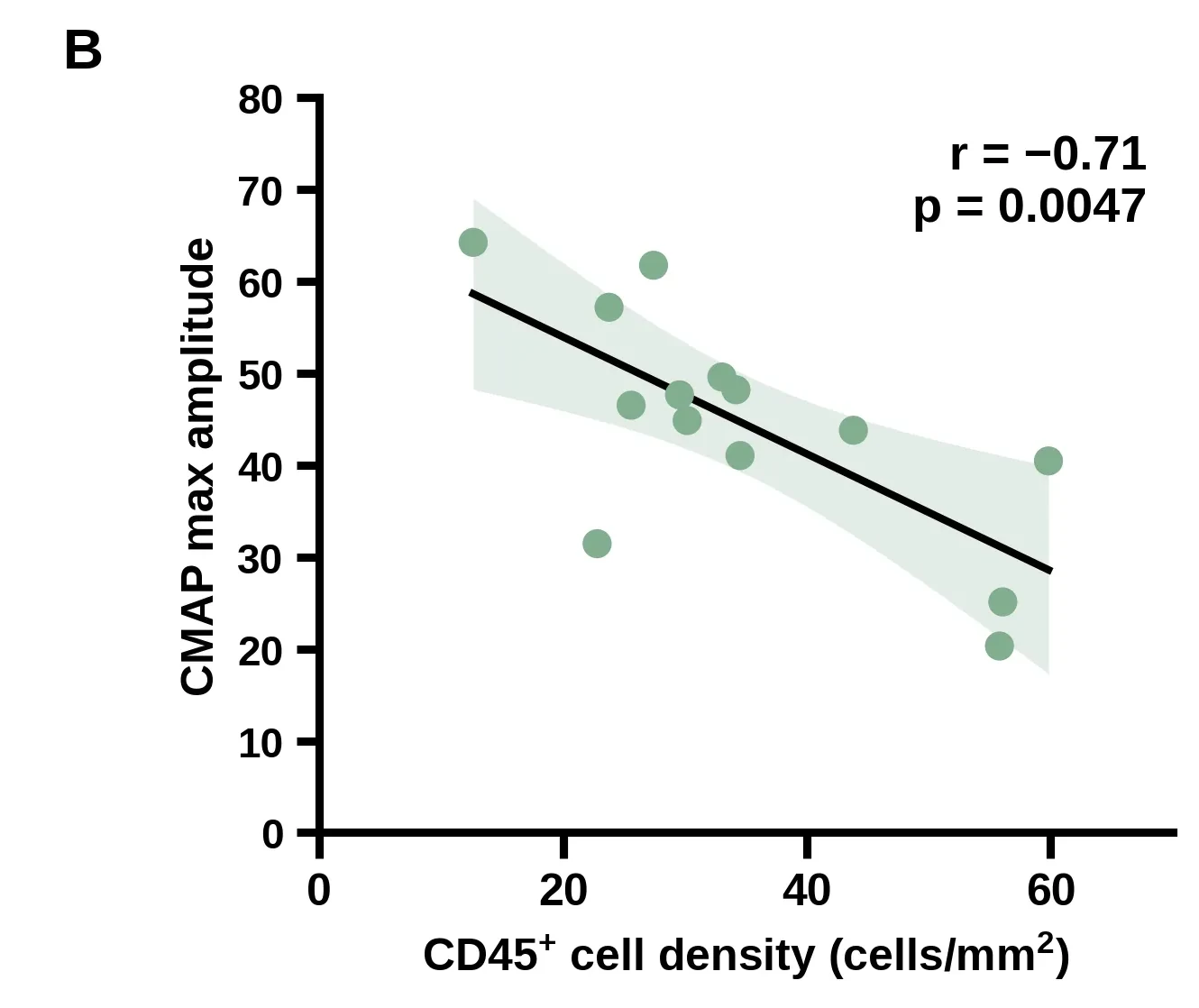
Yes. Across the 14 Low Dox rNLS8 mice, CD45⁺ cell density correlated positively with the density of fully denervated NMJs (Pearson r = 0.81, p = 0.0004) and negatively with CMAP maximum amplitude (r = −0.71, p = 0.0047; Figure 4; Biospective in-house data).
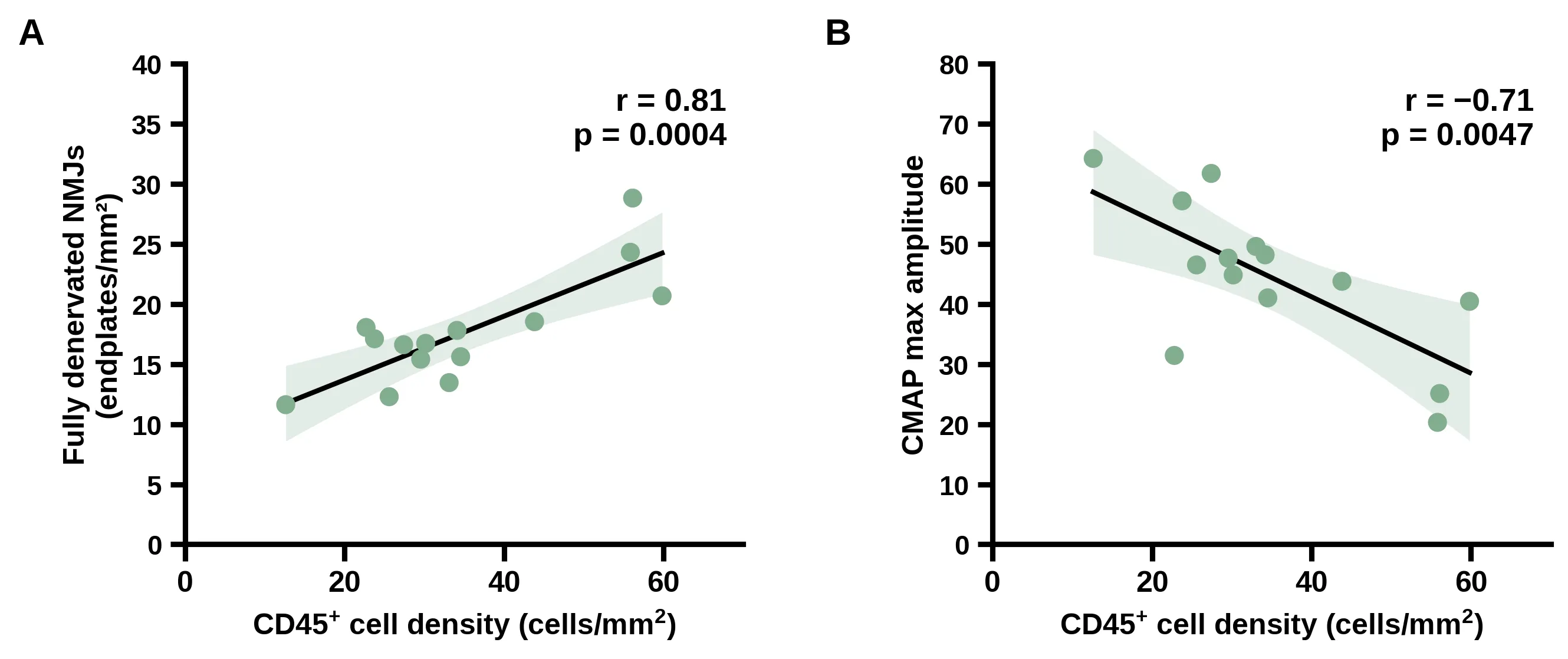
Figure 4. Relationship between gastrocnemius CD45⁺ cell density and (A) the density of fully denervated NMJs (Pearson r = 0.81, p = 0.0004) and (B) CMAP maximum amplitude (Pearson r = −0.71, p = 0.0047) in Low Dox rNLS8 mice; each point is one animal, the solid line is the linear fit and the shaded band its 95% confidence interval; r and p are the values reported from the original analysis. Redrawn in the house palette from Biospective in-house data (study ID to be confirmed); per-animal values were digitized from the original Prism figures in the Interactive Presentation.
These are within-group correlations: they compare Low Dox animals with one another, not Low Dox with controls. They therefore show that inflammatory burden tracks the severity of neuromuscular disease in individual animals, rather than simply marking genotype. Squaring the coefficients (r² ≈ 0.66 and 0.50), CD45⁺ density is statistically associated with roughly two-thirds of the between-animal variance in fully denervated NMJs and about half of the variance in CMAP amplitude (Biospective in-house analysis). Linking a histological immune measure to an electrophysiological readout of functionally innervated muscle fibres is what gives the infiltrate its functional meaning.
Correlation does not establish direction. Immune cells may be recruited by denervation to clear debris, or they may contribute to it, or both. Interventional work in other models supports at least a contributory role: local antibody treatment that reduced leukocyte infiltration also lessened NMJ denervation in hTDP-43 mice (Nógrádi, 2025), deleting C5aR1 in SOD1 mice reduced macrophage recruitment and denervation (Wang, 2017), and masitinib reduced muscle mast cell and macrophage infiltration and lessened denervation in SOD1G93A rats (Trias, 2017). The Low Dox model, with its slower progression, is well suited to testing the same causal question for TDP-43-driven disease.
ONE COHORT, MANY READOUTS
Connect muscle immunology to muscle function
The correlations above were possible because every animal contributed both tissue and electrophysiology. Biospective routinely pairs muscle histology with longitudinal in vivo CT of hindlimb muscle volume, CMAP and grip strength, so you can see whether a change in the infiltrate translates into preserved muscle and function.
Which immune cells make up the muscle infiltrate?
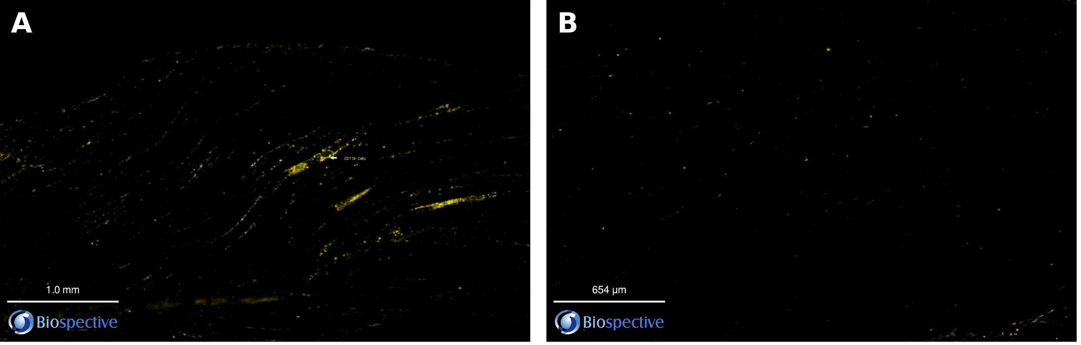
CD11b⁺ myeloid cells, most likely macrophages and related myeloid-lineage cells, appear to make up the bulk of the infiltrate, because CD11b staining on a section consecutive to the CD45-stained section closely mirrored the CD45⁺ distribution in Low Dox muscle and was sparse in tTA controls (Figure 5; Biospective in-house data).
 in gastrocnemius sections from a Low Dox rNLS8 mouse and a tTA control mouse.webp)
Figure 5. CD11b⁺ myeloid cells (yellow) in gastrocnemius sections from (A) a Low Dox rNLS8 mouse (scale bar 1.0 mm), on a section consecutive to the CD45-stained section, and (B) a tTA control mouse (scale bar 654 µm). Panels are still frames captured from the Interactive Presentation viewer at different magnifications and are shown for qualitative comparison only. Biospective in-house data (study ID to be confirmed).
A predominantly myeloid infiltrate is consistent with the macrophage-rich muscle pathology reported in ALS patient biopsies and TDP-43 mice (Nógrádi, 2025) and with macrophage recruitment to denervated muscle in SOD1 models (Van Dyke, 2016; Wang, 2017). Three limitations shape how far this observation can be taken:
CD11b is also expressed by monocytes, neutrophils and some natural killer cells, so it does not by itself identify macrophages.
CD11b is largely absent from conventional T and B lymphocytes, which could contribute to the CD45⁺ population and remain to be quantified.
The CD11b comparison shown here is qualitative; per-animal CD11b density and its relationship to denervation have not yet been quantified.
Adding macrophage markers (F4/80 or CD68), polarization markers that separate pro-inflammatory from pro-regenerative states, Ly6G for neutrophils, CD3 for T cells and CCL2 itself would resolve these questions (the same morphology and phenotyping toolkit we apply to microglia in brain tissue), and matters for interpretation because pro-regenerative macrophages have been associated with slower progression (Margotta, 2023) while mast cells and neutrophils have been implicated in motor axon degeneration (Trias, 2018).
SERVICE
Myeloid & microglia phenotyping
Cell density and morphology classification to separate activated from homeostatic states.
SERVICE
Spatial biology
Map where immune cells sit relative to individual endplates, not just how many there are.
SERVICE
Cytokines & chemokines
Ultrasensitive multiplex immunoassays for CCL2, IL-1β, TNF-α and more.
SERVICE
Myeloid & microglia phenotyping
Cell density and morphology classification to separate activated from homeostatic states.
SERVICE
Spatial biology
Map where immune cells sit relative to individual endplates, not just how many there are.
SERVICE
Cytokines & chemokines
Ultrasensitive multiplex immunoassays for CCL2, IL-1β, TNF-α and more.
How does muscle inflammation in Low Dox rNLS8 mice relate to human ALS?
The muscle phenotype in Low Dox rNLS8 mice mirrors findings in people with ALS, whose muscle shows leukocyte, macrophage, mast cell and neutrophil infiltrates near motor endplates together with endplate denervation and progressive loss of electrophysiological measures of motor units (Bruneteau, 2015; Neuwirth, 2015; Trias, 2018; Nógrádi, 2025).
Correspondence between readouts in the Low Dox rNLS8 mouse and observations or measures in human ALS.
A TDP-43 context for muscle immunology.
Cytoplasmic TDP-43 aggregates are present in nearly all people with ALS, whereas SOD1 mutations account for only a small fraction of cases (Hardiman, 2017; Lépine, 2022). Much of the mechanistic literature on muscle inflammation, however, comes from SOD1 rodents (Van Dyke, 2016; Trias, 2017; Wang, 2017). The Low Dox rNLS8 model provides a TDP-43-driven setting in which the same immune readouts can be measured, complementing recent work in other TDP-43 lines (Nógrádi, 2025).
Clinically aligned functional endpoints.
CMAP amplitude is recorded in the clinic with the same principle used in mice, and derived measures such as the Motor Unit Number Index decline measurably over months in multicentre ALS cohorts, even in muscles that are not yet clinically weak (Neuwirth, 2015; Neuwirth, 2017). Pairing a tissue-level immune measure with CMAP therefore gives sponsors a preclinical readout that maps onto an electrophysiological measure already used in ALS clinical research.
Immunomodulation has reached ALS trials.
Peripheral immune targeting is not hypothetical. Masitinib, a tyrosine kinase inhibitor with activity on mast cells and macrophages, reduced muscle immune infiltration and denervation in SOD1 rodents (Trias, 2017; Harrison & Rafuse, 2020) and was reported to slow functional decline in a prespecified subgroup of a randomized ALS trial (Mora, 2020). Modifying peripheral macrophages has also extended survival in ALS mice (Chiot, 2020). A TDP-43 model with a measurable muscle infiltrate offers a way to test such mechanisms in the pathology that most patients share.
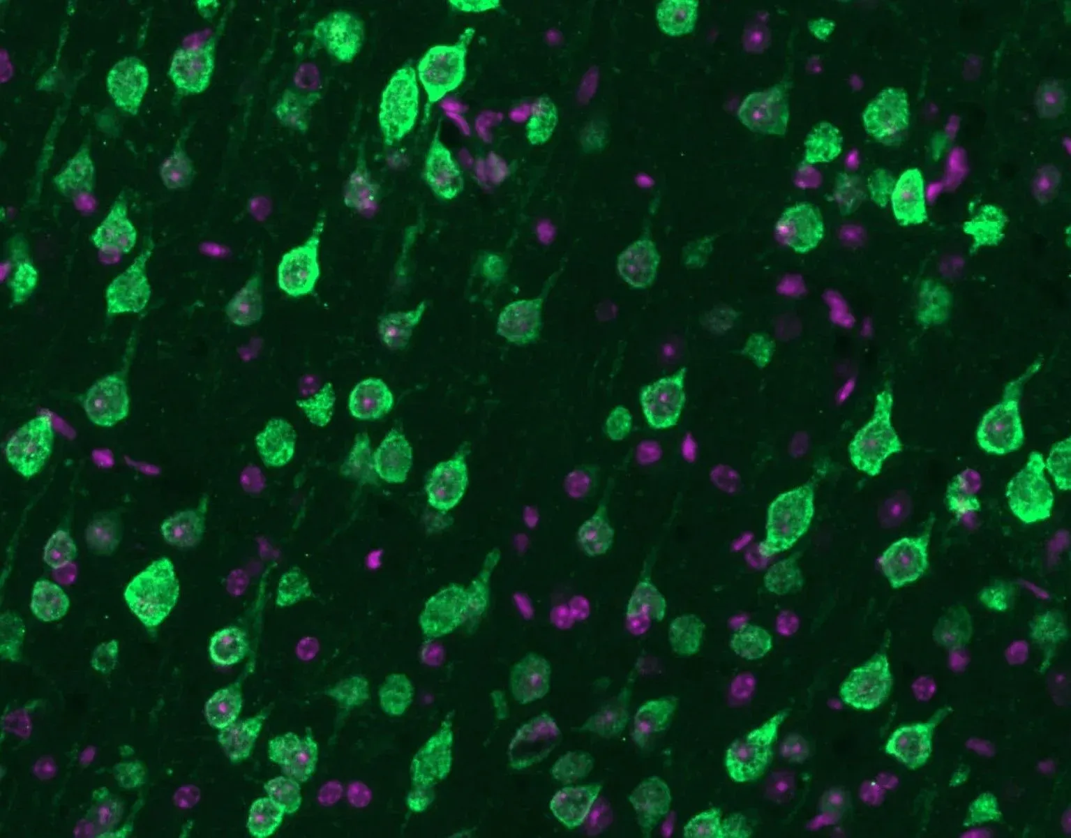
Mislocalization of TDP-43 (green) from the nucleus to the cytoplasm along with nuclei (DAPI) (violet).
CLINICALLY RELEVANT PATHOLOGY
Built on the TDP-43 pathology that most ALS patients share
Cytoplasmic mislocalization of TDP-43, illustrated here, is the defining lesion in nearly all ALS cases (Lépine, 2022). Testing immunomodulatory or neuroprotective candidates in a TDP-43-driven model aligns your preclinical data with the dominant patient population.
How can muscle inflammation be used as an endpoint in preclinical ALS efficacy studies?
Muscle inflammation can be used as a mechanism-of-action and target-engagement endpoint for immunomodulatory ALS therapeutics, and as a disease-severity measure for any therapeutic class, when it is quantified in the same animals as NMJ innervation and CMAP.
Mechanisms this endpoint can read out.
Chemokine-receptor blockade, for example of the CCL2–CCR2 axis (Nógrádi, 2025)
Complement inhibition at C5a–C5aR1 (Wang, 2017)
Tyrosine kinase inhibitors acting on mast cells and macrophages (Trias, 2017; Harrison & Rafuse, 2020)
Macrophage-targeted delivery, such as dendrimer conjugates that localize to activated muscle macrophages (Tallon, 2022)
Stromal and cytokine pathways in denervated muscle, such as fibro-adipogenic progenitor STAT3–IL-6 signalling (Madaro, 2018)
Strategies that reprogramme peripheral macrophages rather than deplete them (Chiot, 2020)
An integrated, same-animal endpoint package.
Because CD45⁺ density, NMJ innervation classes, presynaptic complexity and CMAP come from the same mice, a drug-induced reduction in the infiltrate can be checked directly for downstream benefit to synapses and muscle function, and a drug that rescues NMJs without touching inflammation can be distinguished from one that acts through it. These readouts can be combined with the grip strength, CT muscle volume, plasma and CSF neurofilament light chain (NfL), MRI brain atrophy and brain histopathology endpoints already validated in this model, and can be complemented by cytokine profiling, as described on our TDP-43ΔNLS model page.
Inflammatory burden as a covariate.
Because CD45⁺ density accounted for a large share of between-animal variation in denervation and CMAP in this cohort, it can also be analysed as a covariate or stratification variable in studies of non-immune therapies, helping to separate genuine treatment effects from differences in disease severity among animals (Biospective in-house analysis).
Design considerations we discuss with sponsors.
Dosing window and route: prevention designs starting at induction versus intervention designs starting once denervation is established, using any of our routes of administration.
Positive control: reinstating doxycycline halts transgene expression and allows reinnervation and functional recovery, providing a benchmark for disease modification (Walker, 2015).
Phenotype, not just number: broad myeloid suppression can be counterproductive, since microglia support recovery in this model and pro-regenerative macrophages are associated with slower progression (Spiller, 2018; Margotta, 2023), so polarization markers are recommended for immunomodulatory programmes.
Sample size: in our earlier NMJ dataset from this model, fewer than 15 mice per group were estimated to detect a 25% change in SV2A–α-BTX overlap; equivalent estimates for the CD45⁺ endpoint can be derived from the present cohort (Biospective in-house data).
Sponsors receive whole-section image data, per-animal quantitative metrics, NMJ class maps, with interactive presentations of study sections available on request.
PARTNER WITH BIOSPECTIVE
Ready to test your ALS therapeutic in the Low Dox rNLS8 model?
In-house rNLS8 breeding colony at Biospective for rapid study start-up and large cohorts
Slower Low Dox progression that widens the window for detecting disease modification
Experience with small molecules, antibodies, antisense oligonucleotides, gene therapies and peptides by multiple routes
Muscle inflammation, NMJ innervation, CMAP, CT, grip strength, NfL, MRI and brain pathology from the same animals
Whole-slide, interactive image delivery like the presentation on this page
Summary
In the gastrocnemius muscle of Biospective's Low Dox rNLS8 mice, CD45⁺ immune cells are roughly twice as dense as in tTA controls, appear to be predominantly CD11b⁺ myeloid cells, and accompany marked NMJ denervation and axonal pathology (Biospective in-house data). Within Low Dox animals, inflammatory burden tracks both fully denervated NMJs and CMAP amplitude, linking the infiltrate to structural and functional neuromuscular decline. These findings parallel the leukocyte and macrophage infiltrates now documented in ALS patient muscle (Nógrádi, 2025) and position muscle inflammation as a translational, mechanistically informative endpoint for preclinical ALS therapeutics. Explore our full portfolio of ALS mouse models and our Microglia, Astrocytes & Neurodegeneration Initiative.
FAQs
FAQs
References
Keywords
Related Content
Related Content
RESOURCE
Neuromuscular Junction (NMJ) Morphology & ALS Models
How NMJs are affected in ALS and the methods used to measure morphological change.
RESOURCE
TDP-43ΔNLS (rNLS8) Mice for ALS Drug Development
Using the ΔNLS model for preclinical therapeutic studies, from induction to endpoints.
RESOURCE
A Guide to ALS Models for Drug Discovery
Strengths and limits of mouse and rat ALS models for testing therapeutics.
RESOURCE
Neuromuscular Junction (NMJ) Morphology & ALS Models
How NMJs are affected in ALS and the methods used to measure morphological change.
RESOURCE
TDP-43ΔNLS (rNLS8) Mice for ALS Drug Development
Using the ΔNLS model for preclinical therapeutic studies, from induction to endpoints.
RESOURCE
A Guide to ALS Models for Drug Discovery
Strengths and limits of mouse and rat ALS models for testing therapeutics.