


6-OHDA Mice vs. AAV-α-Synuclein Mice for PD Programs
Which Parkinson's mouse model fits your program: pathology, time course, variability and readouts compared.
 What are 6-OHDA mice and AAV-α-synuclein mice?
- How do the two models differ in α-synuclein pathology and target engagement?
- How do the two models differ in progression, variability and reproducibility?
- How does model choice affect study timelines and cohort size?
- What endpoints can Biospective measure in the AAV-A53T-α-synuclein model?
- Which model should you choose for your Parkinson's program?
Summary
In brief: 6-OHDA mice and AAV-α-synuclein mice address different research questions. The 6-OHDA lesion produces a rapid oxidative insult that denervates the nigrostriatal pathway without reproducing the characteristic α-synuclein aggregation seen in Parkinson’s disease, making it a useful platform for symptomatic and dopaminergic-rescue studies. In contrast, AAV-mediated expression of human A53T α-synuclein induces phosphorylated α-synuclein pathology in nigral neurons alongside degeneration of the nigrostriatal pathway. In a head-to-head comparison in rats, AAV-A53T reached motor impairment equivalent to 6-OHDA, but with less cell loss and persistent axonal pathology not observed in the toxin lesions (Decressac, 2012). Because the pharmacodynamic evaluation of α-synuclein-directed therapies relies on α-synuclein-related measures (Naki, 2026), the 6-OHDA model is not well suited for assessing target engagement by these therapies. Biospective's AAV-A53T mouse model, in contrast, produces motor deficits by Week 5, accompanied by pSer129 pathology, dopaminergic loss, glial activation, 7T MRI-detected atrophy, and changes in CSF neurofilament light. Together, these features make it a strong option for evaluating disease-modifying therapies targeting α-synuclein.
Video 1: Human Ser129 pathology, the two induction routes, the analytes used to evaluate α-synuclein-directed therapies, the clinical record, and the readouts available in the 6-OHDA and AAV-A53T-α-synuclein mouse models.
What are 6-OHDA mice and AAV-α-synuclein mice?
6-OHDA at a glance
A brief overview of the 6-OHDA model of Parkinson’s disease.
Where the toxin goes
6-OHDA cannot cross the blood-brain barrier (BBB), so it is injected stereotaxically into the medial forebrain bundle (MFB), the substantia nigra (SN), or the striatum. The site chosen sets how fast and how complete the dopaminergic loss is.
What it produces
Dose-graded loss of nigral dopaminergic neurons and striatal terminals, motor deficits, several non-motor features, and astrocytic and microglial activation that persists for weeks.
What it does not produce
The model does not reproduce the characteristic α-synuclein aggregation and Lewy-like pathology of Parkinson’s disease. Consequently, it is not well suited to evaluating therapies that target pathological α-synuclein.
Where the toxin goes
6-OHDA cannot cross the blood-brain barrier (BBB), so it is injected stereotaxically into the medial forebrain bundle (MFB), the substantia nigra (SN), or the striatum. The site chosen sets how fast and how complete the dopaminergic loss is.
What it produces
Dose-graded loss of nigral dopaminergic neurons and striatal terminals, motor deficits, several non-motor features, and astrocytic and microglial activation that persists for weeks.
What it does not produce
The model does not reproduce the characteristic α-synuclein aggregation and Lewy-like pathology of Parkinson’s disease. Consequently, it is not well suited to evaluating therapies that target pathological α-synuclein.
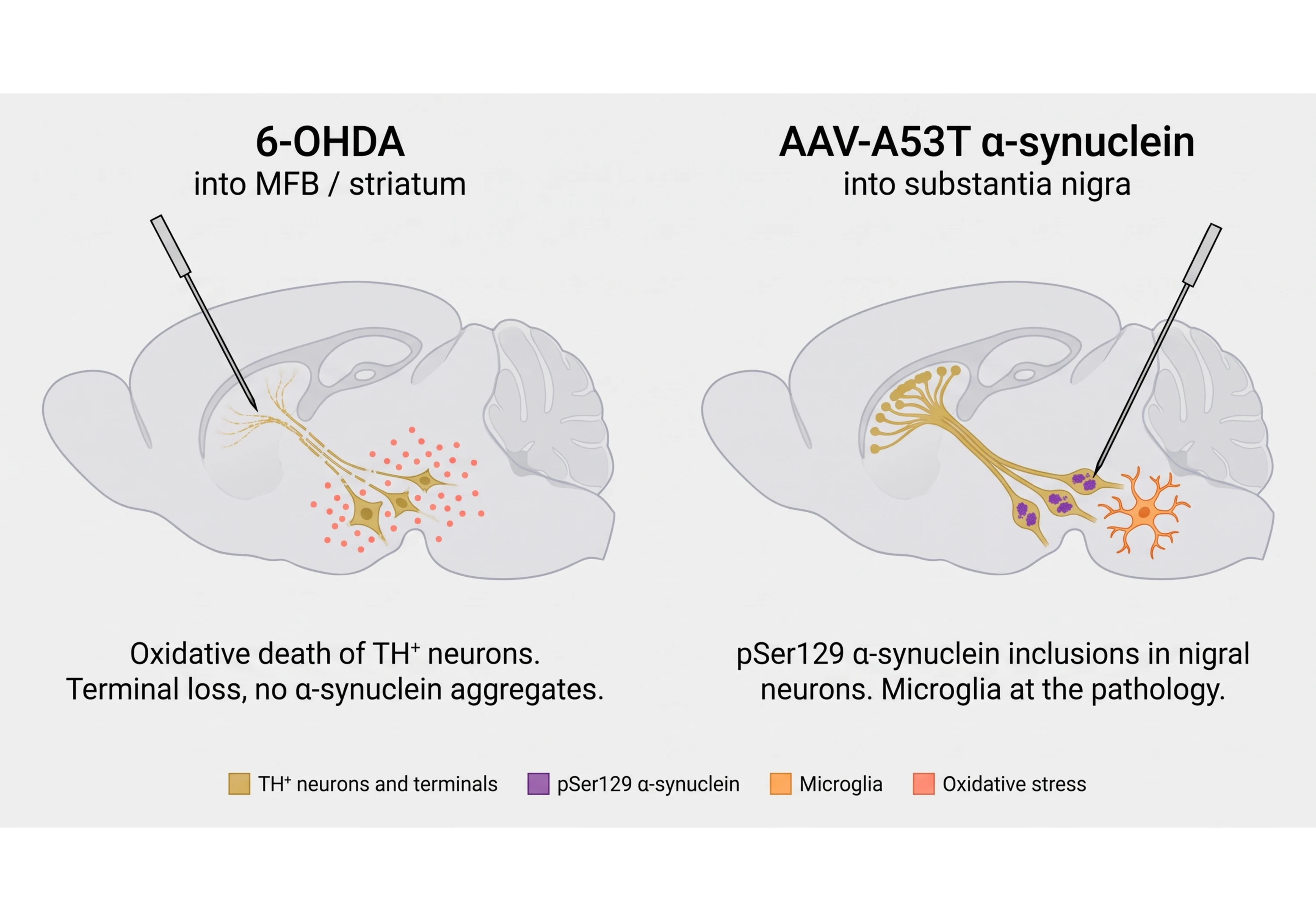
6-OHDA mice are animals given a stereotaxic injection of the catecholaminergic neurotoxin 6-hydroxydopamine to destroy nigrostriatal dopamine neurons, while AAV-α-synuclein mice are given an adeno-associated viral (AAV) vector that drives the overexpression of human α-synuclein in these neurons, which can lead to their degeneration. In other words, 6-OHDA causes dopaminergic neurodegeneration through oxidative stress, whereas AAV-mediated α-synuclein overexpression induces α-synuclein-associated pathology. That difference determines which therapeutic questions each model can answer.
The 6-OHDA lesion
6-OHDA does not cross the blood-brain barrier (BBB), so it is delivered stereotactically into the medial forebrain bundle (MFB), the substantia nigra (SN), or the striatum. Once inside catecholaminergic terminals, it drives cytosolic dopamine accumulation and auto-oxidation, resulting in oxidative stress and neuronal death. A recent synthesis proposes vesicular monoamine transporter 2 (VMAT2) as a shared node in this cascade, although this is presented as a unifying hypothesis rather than an experimentally tested mechanism (Zhu, 2025).
The lesion is graded by dose: in mice, unilateral medial forebrain bundle injection at 0.5, 1, and 2 µg produced dose-dependent nigral TH⁺ cell loss and striatal fiber loss, along with sustained astrocytic and microglial activation at seven weeks (Cui, 2024). Intrastriatal lesioning in male mice can reproduce motor deficits, cognitive impairment, and gastrointestinal dysfunction within a four-week protocol. However, olfactory dysfunction and anxiety-like behavior were not reproduced, and catecholamine disruption extended beyond the nigrostriatal pathway into mesocortical and mesolimbic systems (Braun, 2025).
What the lesion does not do is induce Lewy-like pathology. A PRISMA systematic review of the 6-OHDA literature found that the model does not reproduce the α-synuclein and Lewy body pathology of Parkinson's disease, despite effectively degenerating the nigrostriatal dopaminergic tract (Prates-Rodrigues, 2025). The mechanistic literature treats α-synuclein only as a hypothesized upstream modulator of the same oxidative node, not as a product of the lesion (Zhu, 2025).
The AAV-α-synuclein model
AAV delivery of human α-synuclein into the substantia nigra produces a progressive lesion that can begin with changes at the axon terminals. In rats, motor impairment appeared at four and six weeks after AAV-A53T injection and correlated with striatal terminal loss rather than nigral cell counts, suggesting that the behavioral phenotype is closely associated with denervation of the target field (Musacchio, 2022).
Severity is titer-dependent, with distinct thresholds: an intermediate AAV1/2-A53T titer produced paw-use asymmetry and reduced striatal TH at three weeks, progressing to nigral cell loss and dopamine depletion by six weeks. A more dilute preparation produced deficits that compensated between three and six weeks, while an excessive titer caused non-specific toxicity that was also observed in GFP control animals (Koprich, 2011). Overabundance of α-synuclein can also impair dopamine release beyond what would be expected from cell loss alone. In rats overexpressing wild-type human α-synuclein, reductions in striatal dopamine release exceeded the loss of dopaminergic neurons, axonal fibers, and total dopamine, and were accompanied by decreased vesicle density and fewer synaptic contacts (Gaugler, 2012).
Biospective's implementation is a unilateral injection of AAV-human-A53T-α-synuclein into the substantia nigra of 12 week-old C57BL/6 mice, with the contralateral hemisphere and a control group serving as comparators. The procedure is performed through our stereotaxic surgery service. Robust motor deficits are present by Week 5 across the cylinder test, rotarod, and tail suspension swing test. These changes are accompanied by reduced nigral TH⁺ neurons, near-complete loss of ipsilateral striatal TH⁺ terminals, reduced striatal dopamine, pSer129 α-synuclein pathology, microglial and astrocytic activation, 7T MRI atrophy, and elevated CSF neurofilament light (NfL). Together, these findings provide a range of behavioral, histological, imaging, and fluid biomarker readouts for evaluating therapeutic effects in this model. Full details are available on our AAV synuclein models page, alongside the rest of our Parkinson's disease rodent models.
Match the model to your mechanism
A symptomatic compound needs nigrostriatal function. An α-synuclein-directed compound needs a model with relevant α-synuclein pathology and pharmacodynamic readouts. See what the AAV-A53T model measures, and decide which route your program is on.
Match the model to your mechanism
A symptomatic compound needs nigrostriatal function. An α-synuclein-directed compound needs a model with relevant α-synuclein pathology and pharmacodynamic readouts. See what the AAV-A53T model measures, and decide which route your program is on.
Table 1. 6-OHDA mice and AAV-A53T-α-synuclein mice compared on the features that matter for preclinical drug development.
Note: "Not replicated" follows the systematic-review finding that the 6-OHDA model does not reproduce α-synuclein and Lewy body pathology; "None available" follows from it rather than from a formal negative result. Rows comparing cell loss and axonal pathology between the models are from a same-laboratory comparison in rats, and transfer to mice as a principle rather than as a measurement. Rows describing Biospective's mouse model are in-house data.
Sources: Braun, 2025; Cui, 2024; Decressac, 2012; Gaugler, 2012; Koprich, 2011; Musacchio, 2022; Prates-Rodrigues, 2025; Zhu, 2025.
Induction Routes. Both induction routes converge on nigrostriatal dopaminergic loss and motor deficits, but only the AAV-A53T route passes through pSer129 α-synuclein pathology.
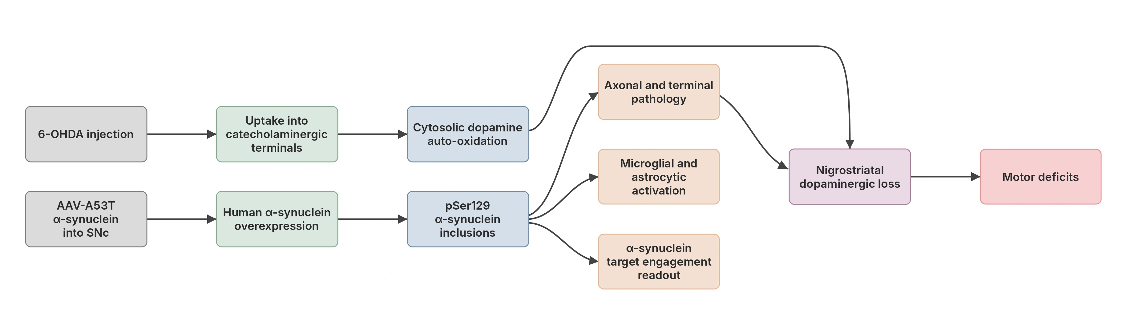
Schematic illustration of the two induction routes. Both converge on nigrostriatal dopaminergic loss and motor deficits, but only the AAV-A53T α-synuclein route passes through pSer129 inclusions, which are associated with glial activation and provide a target engagement readout.
How do the two models differ in α-synuclein pathology and target engagement?
The two models differ substantially here: AAV-α-synuclein mice express human α-synuclein and can develop associated pathological protein species, whereas 6-OHDA mice do not reproduce the characteristic α-synuclein pathology of Parkinson’s disease (PD). For a symptomatic program, this distinction may be less relevant. For a disease-modification program, however, it can determine whether the model provides an appropriate readout for assessing target engagement.
What the human pathology is
Phosphorylation at serine 129 is the dominant pathological modification of α-synuclein in human Lewy body disease. Mass spectrometry of synucleinopathy lesions showed that Ser129 is selectively and extensively phosphorylated in Parkinson's disease, dementia with Lewy bodies, and multiple system atrophy (MSA), and that Ser129 phosphorylation is associated with fibril formation (Fujiwara, 2002). An unbiased inventory of α-synuclein forms in Lewy bodies confirmed that phosphorylation at Ser-129 is the predominant modification, with the same pattern in SNCA A53T familial Parkinson's disease and in multiple system atrophy (MSA), pointing to a shared pathway across genetic and sporadic disease (Anderson, 2006).
The AAV-A53T model reproduces pSer129 α-synuclein pathology in the substantia nigra, whereas the 6-OHDA lesion does not reproduce this characteristic pathological feature.
Why target engagement decides the model
Every active α-synuclein-directed modality is evaluated using pharmacodynamic measures related to its α-synuclein target, rather than dopaminergic survival alone. A brain-region-resolved study of an antisense oligonucleotide (ASO) against the human SNCA 3′UTR in BAC-hSNCA rats measured human SNCA mRNA and soluble and detergent-insoluble α-synuclein protein as its primary pharmacodynamic endpoints, finding regionally graded suppression strongest in the olfactory bulb and striatum at a stage preceding overt dopamine depletion (Naki, 2026). These measurements illustrate the types of target-related readouts that can be used in an α-synuclein model. A 6-OHDA lesion, which does not reproduce human α-synuclein pathology or express a human SNCA transgene, is not suitable for evaluating these specific pharmacodynamic endpoints. It can register neuroprotection, but cannot establish whether an α-synuclein-directed drug has engaged its intended target using these measures.
The clinical record makes the importance of this distinction clear. In the PASADENA Phase 2 trial, 316 participants with early Parkinson's disease received prasinezumab 1500 mg, 4500 mg, or placebo for 52 weeks; the primary MDS-UPDRS endpoint was not met, with differences from placebo of −2.0 (80% CI −4.2 to 0.2; P = 0.24) and −0.6 (80% CI −2.8 to 1.6; P = 0.72), and no substantial difference on DaT-SPECT (Pagano, 2022). The larger PADOVA Phase 2b study in 586 participants also did not meet its primary endpoint, with a hazard ratio of 0.84 (95% CI 0.69–1.01; p = 0.066) for time to confirmed motor progression and a median of 61.1 versus 49.7 weeks (Nikolcheva, 2026). Without a suitable pharmacodynamic measure of target engagement, it can be difficult to distinguish an ineffective therapeutic target from insufficient drug exposure or target engagement. That distinction is important to address during preclinical development, using a model and readouts appropriate to the therapeutic target.
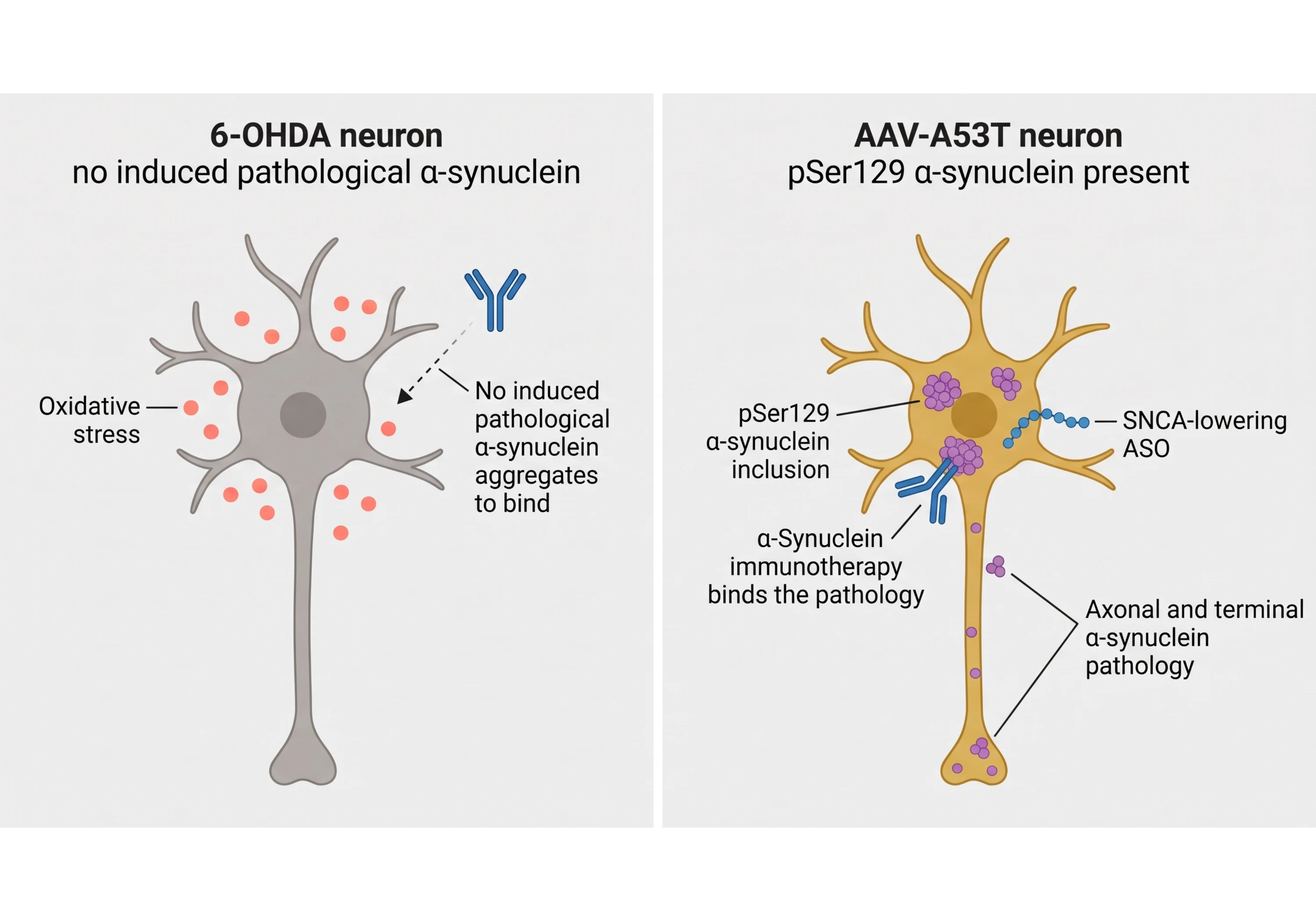
Target engagement. The same comparison in a single nigral neuron: a 6-OHDA neuron does not provide the experimentally induced human α-synuclein pathology needed for direct target-engagement assessment, while an AAV-A53T neuron carries pSer129 α-synuclein in the soma, axon and terminals.
Translational limitations of toxin models
Two reviews highlight limitations in the translation of preclinical neuroprotective findings to clinical disease-modifying therapies. A narrative review of two decades of disease-modification trials contrasts the historical success of translating preclinical work into symptomatic therapies with an accumulation of failures for disease-modifying interventions that had worked in preclinical models (Mari, 2022). A review of neuroprotection trials attributes the failure to translate primarily to an incomplete understanding of Parkinson's pathogenesis and to excessive reliance on data from toxin-based animal models when selecting agents for clinical trials, compounded by the absence of validated progression biomarkers (Athauda, 2014).
None of this makes 6-OHDA a bad model. Rather, it highlights that 6-OHDA is not the right model for every disease-modifying therapeutic program, particularly those targeting α-synuclein pathology.
How do the two models differ in progression, variability and reproducibility?
The 6-OHDA model produces a faster, more abrupt lesion; the AAV-α-synuclein lesion is generally slower, can begin with changes at the axon terminals, and is influenced by vector titer. Both are controllable, but they are controlled by different factors, and the published variability profiles differ.
Speed and shape of the lesion
How fast a 6-OHDA lesion develops depends on where the toxin is delivered. Injection into the medial forebrain bundle (MFB) produces near-complete dopaminergic destruction rapidly, whereas intrastriatal injection primarily affects terminals initially, followed by degeneration of nigral cell bodies over the following weeks. This is why intrastriatal protocols are often used to model a more progressive pattern of dopaminergic degeneration. Either way, the insult is delivered once, at surgery, and is not ongoing by the time most behavioral testing begins.
AAV-α-synuclein pathology develops over several weeks instead: paw-use asymmetry and reduced striatal TH appear around three weeks at an intermediate titer and progress to nigral cell loss and dopamine depletion by six weeks (Koprich, 2011), with motor impairment at four and six weeks tracking striatal terminal loss rather than nigral cell counts (Musacchio, 2022). A longitudinal rat characterization found nigrostriatal pathology and behavioral deficits within a four-week period after rAAV2/7 α-synuclein, with the phenotype reversible by L-DOPA and followable non-invasively by PET and MR spectroscopy (Van der Perren, 2015).
Progressive pathology can provide a therapeutic window, allowing a compound to be dosed before or during degeneration rather than only after it.
The lesion burden associated with a given deficit
This is where the only direct head-to-head comparison is informative. Testing AAV-α-synuclein rats against both partial intrastriatal and full medial forebrain bundle 6-OHDA lesions in the same laboratory, with longitudinal behavior and matched histology, the authors found differences in the relationship between motor impairment and dopaminergic degeneration across models: α-synuclein animals showed less cell and terminal loss for an equivalent level of motor abnormality, and prominent persistent axonal pathology appeared only in the α-synuclein model. The authors concluded that the α-synuclein model more closely replicated aspects of human pathology than either toxin lesion (Decressac, 2012). This provides a useful comparison, although the findings were obtained in rats.
Where each model's variability comes from
Variability in the 6-OHDA model is largely procedural. Authors characterizing the intrastriatal mouse model note that it is not well characterized, with variable dosages, injection sites, and timeframes across the literature (Braun, 2025). Dose does not buy unlimited dynamic range either: in mouse medial forebrain bundle lesions, 2 µg produced more neuronal loss than 1 µg but no further behavioral impairment, so the behavioral readout reached a ceiling before the histological readout (Cui, 2024).
AAV variability is largely a titer and vector-control problem. Too dilute a preparation gives deficits that compensate away between three and six weeks; too concentrated a preparation causes non-specific toxicity visible even in GFP control animals (Koprich, 2011). Biospective's AAV-A53T studies can report TH alongside striatal dopamine, pSer129 burden, glial morphometry, structural MRI, and CSF neurofilament light (NfL), so no conclusion rests on a single marker. Titer is a design variable rather than a fixed setting: cylinder test and tail suspension swing test deficits have been observed to vary across low, moderate, and high titers in our hands, which can help position a study for prevention or rescue approaches.
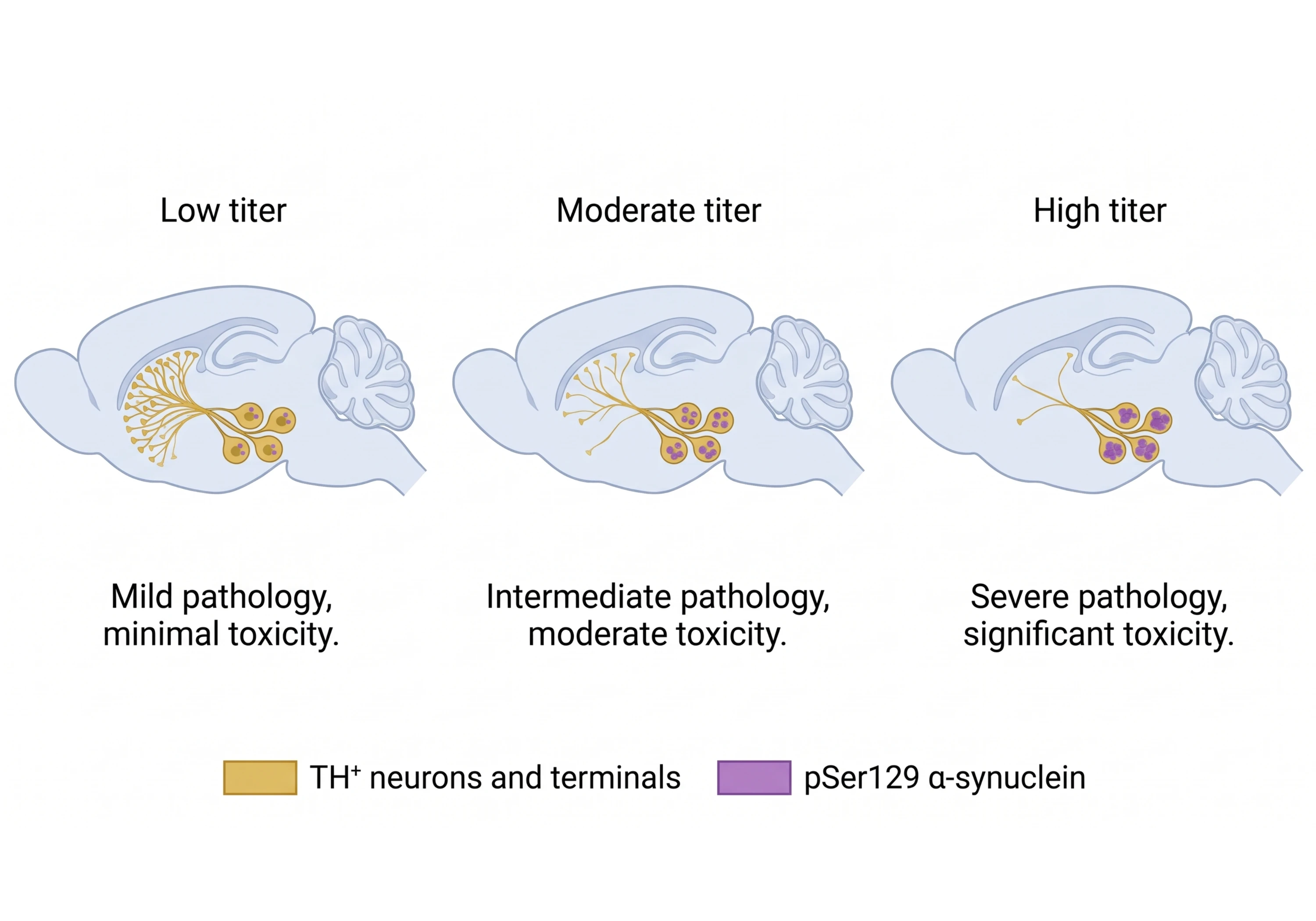
Titer grading. AAV titer influences lesion severity. Deficits can compensate away at low titer and may reach a behavioral floor at high titer; an intermediate titer gives the largest effect size.
What this means for choosing a preclinical Parkinson's model
Neither model is intrinsically more reproducible; rather, their major sources of variability differ. A preclinical Parkinson's model built on 6-OHDA is reproducible to the extent that target, dose, and lesion verification are standardized, and a model built on AAV-α-synuclein is reproducible to the extent that titer and vector controls are fixed and endpoints are interpreted together.
How does model choice affect study timelines and cohort size?
Model choice changes three things in a study plan: how long you wait for a readout, how many endpoints one animal can provide, and how much of the variance is up to the investigator to control.
Time to readout
Both models provide a motor readout within a few weeks of surgery. In Biospective's AAV-A53T mouse model, robust deficits across the cylinder test, rotarod, and tail suspension swing test are present at Week 5. In practice, this allows dosing to begin shortly after injection, with terminal collection scheduled once the phenotype is established. Published 6-OHDA protocols can be shorter — the intrastriatal mouse protocol runs to four weeks (Braun, 2025) — so if speed alone were the criterion, 6-OHDA would have an advantage.
Speed alone is rarely the criterion. The relevant comparison is time to a readout the program can act on, and a model that does not reproduce α-synuclein pathology cannot provide pathology-based readouts for an α-synuclein-directed compound, regardless of how quickly the lesion develops.
What drives cohort size
Cohort size is driven by effect size and residual variance, and no published study quantifies how lesion variability differs between 6-OHDA and AAV-α-synuclein mice on matched endpoints. What the literature does establish is where the variance comes from. A PRISMA systematic review of 135 6-OHDA rodent studies found significant variability in brain target, toxin dosage, lesion verification strategy, and behavioral assessment reporting, and identified this heterogeneity as a direct obstacle to reproducibility and cross-study comparability, calling for standardization and consistent evaluation of lesion extent (Prates-Rodrigues, 2025). Standardizing these factors within a study can help reduce procedural variability, regardless of the model selected.
Three design choices can influence study design and cohort size, and all three are available in the AAV model:
Graded severity. Titer sets the depth of the lesion, with compensation at the low end and non-specific toxicity at the top end as the boundaries (Koprich, 2011); a study can be positioned where the effect size is expected to be useful rather than simply where the lesion is deepest.
Multiple endpoint classes in the same animal. Behavior, in vivo MRI, histology, and CSF biomarkers provide complementary measures of treatment effects, so a single failed readout does not necessarily determine the outcome of the entire study.
A within-animal comparator. A unilateral lesion leaves the contralateral hemisphere as an internal reference for histological and volumetric measures.
Scope your Parkinson's efficacy study
Behavior, in vivo imaging, histology and fluid biomarkers from one cohort, so a disease-modifying claim rests on measures that agree. Tell us your therapeutic modality, target, and timeline and we will design around them.
Scope your Parkinson's efficacy study
Behavior, in vivo imaging, histology and fluid biomarkers from one cohort, so a disease-modifying claim rests on measures that agree. Tell us your therapeutic modality, target, and timeline and we will design around them.
One caution about the contralateral control
The internal control is useful, but it is not perfectly neutral. Whole-brain voxel-based morphometry after unilateral 6-OHDA lesioning in rats found gray matter volume changes on both sides, strongest in the lesioned hemisphere, but also present contralaterally (Westphal, 2016). For that reason, ipsilateral–contralateral differences should be interpreted alongside a separate control group, rather than using the contralateral hemisphere as the sole control.
How in vivo imaging supports longitudinal assessment
Longitudinal imaging allows changes to be followed over time within the same animal, making it possible to compare preclinical and clinical measures collected over very different timescales. Non-invasive PET and MR spectroscopy followed neurodegeneration in AAV-α-synuclein rats over a reproducible four-week window (Van der Perren, 2015). The human equivalents take years: a meta-analysis of 39 studies in 1,983 patients found neuromelanin-sensitive MRI measures associated with motor impairment (pooled r = −0.406), disease duration (r = −0.426) and Hoehn and Yahr stage (r = −0.336), all P < 0.001, with only four longitudinal studies reporting annual declines of about 5% to 19% (Akande, 2026).
Biospective's 7T magnetic resonance imaging (MRI) shows substantia nigra and caudate-putamen atrophy that scales with AAV titer and aligns with TH⁺ neuron and terminal loss. Although these preclinical structural MRI measures are not equivalent to the neuromelanin-sensitive MRI measures used clinically, both provide longitudinal information about changes in relevant brain regions. This strategy offers a way to assess treatment-related changes in a preclinical study over weeks, alongside clinical imaging findings collected over years.
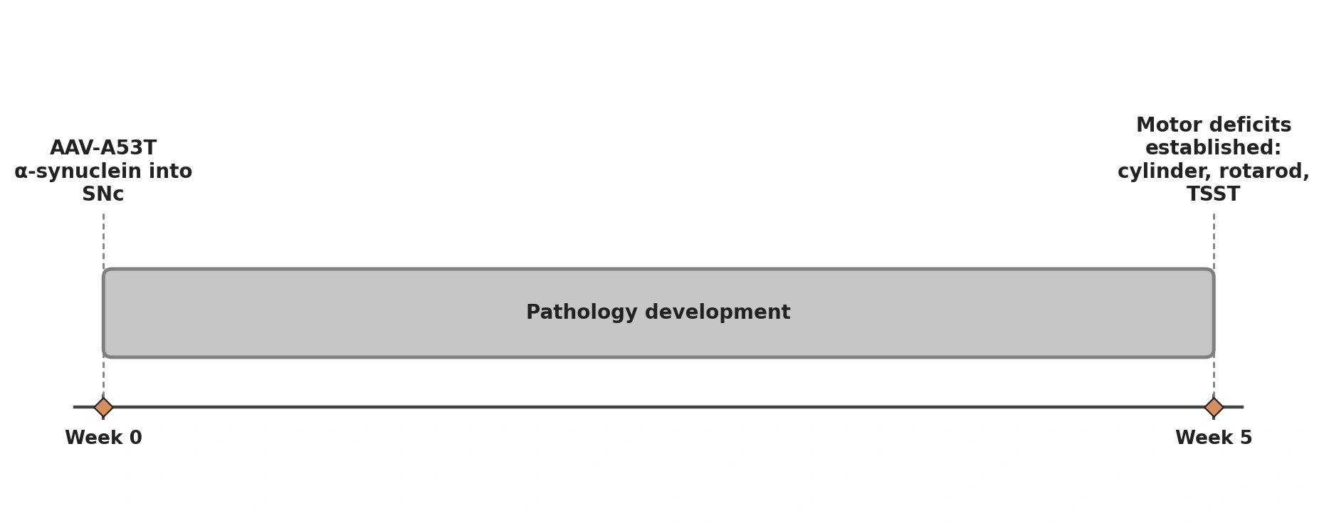
Schematic illustration of the in-life window in Biospective's AAV-A53T α-synuclein mouse model: unilateral injection into the substantia nigra at Week 0, pathology developing over the following weeks, and motor deficits established across the cylinder test, rotarod and tail suspension swing test by Week 5.
What endpoints can Biospective measure in the AAV-A53T-α-synuclein model?
Biospective runs the model end-to-end and reads it out in four endpoint classes from the same mice: behavior, in vivo imaging, tissue analysis, and fluid biochemistry. The point of running them together is that treatment effects can be evaluated across complementary measures rather than relying on any single endpoint.
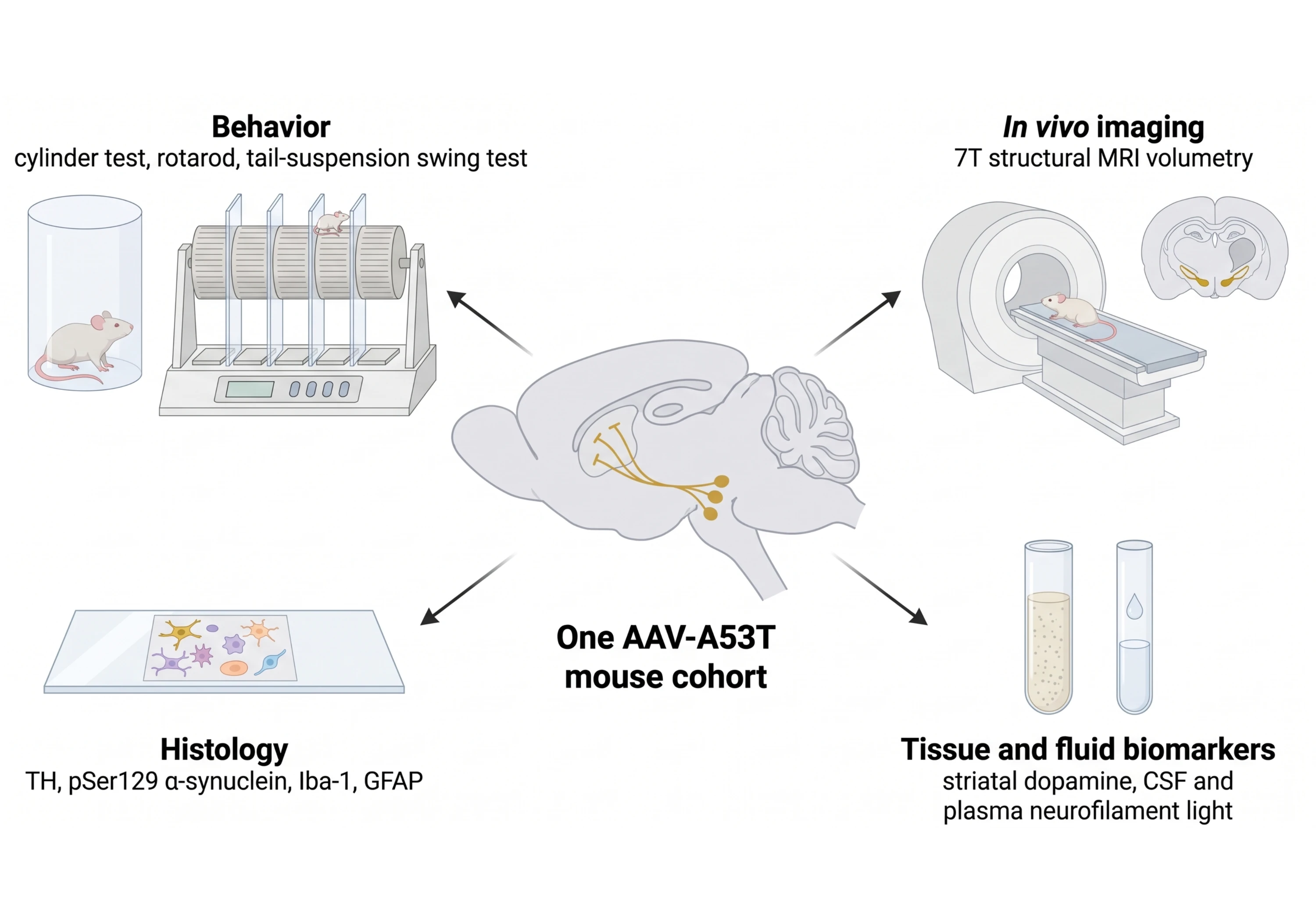
Endpoint classes. Behavior, 7T MRI, multiplex histology, and tissue/fluid biomarkers read out from the same AAV-A53T mice, so a disease-modifying claim rests on measures that agree.
Behavior
Motor function is assessed with the cylinder test for forelimb asymmetry, rotarod for motor coordination and the tail suspension swing test for directional bias, through our motor and sensory function testing. In our hands, all three distinguish AAV-Syn from control animals at Week 5-6, and clasping, cylinder and swing-test deficits vary with low, moderate, and high AAV titers. Because the motor phenotype in this model tracks striatal terminal loss rather than nigral cell counts (Musacchio, 2022), behavior is interpreted as a functional readout associated with nigrostriatal denervation and reported alongside the anatomy, not in place of it.
In vivo imaging
7T structural MRI measures substantia nigra and caudate-putamen volume, which in our data scales with AAV titer and aligns with TH⁺ neuron and terminal loss. Imaging also makes the design longitudinal: each animal is its own baseline, allowing structural changes to be followed over time. In the clinical field, neuromelanin-sensitive MRI measures nigral changes, with reported annual declines of roughly 5% to 19% (Akande, 2026).
Tissue analysis
Multiplex immunofluorescence with automated quantification covers TH⁺ neurons and terminals, pSer129 α-synuclein burden, dopaminergic nuclei, and glial morphology. Microglial and astrocytic responses are quantified by morphology, rather than by stain density alone: Iba-1⁺ density and activated microglia rise in midbrain, SNc and SNr, and GFAP⁺ density and hypertrophic astrocytes rise in midbrain and SNc, with microglia clustering where pSer129 aggregates sit. Our microglia histology and analysis service applies the same pipeline across models.
Glial endpoints are informative. Microglial activation appears early in viral α-synuclein models, alongside motor decline rather than necessarily following it (Kefalakes, 2025), and there is evidence that microglia can contribute to α-synuclein-associated pathology: in a mouse model combining AAV α-synuclein overexpression with intranigral fibrils, removing T cells reduced α-synuclein pathology and microglial activation, while depleting microglia reduced T-cell accumulation and lessened pathology (Ohtake, 2026).
The human counterpart is measurable: in a cohort of newly diagnosed patients, the substantia nigra was the only region where ¹¹C-PK11195 binding was higher in those cognitively impaired at three years than in controls, and baseline nigral binding together with a CSF-to-blood memory B-cell ratio predicted decline, the leading markers reaching AUCs of 0.79 to 0.85 (Scott, 2025).
Fluids and biochemistry
Striatal dopamine is quantified directly from brain homogenates, and CSF and plasma are collected for fluid and cellular biomarkers, including our neurofilament light chain (NfL) assay. CSF NfL is elevated in AAV-Syn mice in our data.
Two caveats belong with that number. NfL is a validated but non-specific marker: a Bayesian network meta-analysis of 78 studies and 13,120 participants found CSF and blood NfL raised in Parkinson's disease relative to controls but higher still in atypical parkinsonism, with CSF NfL in multiple system atrophy (MSA) exceeding Parkinson's by an SMD of 1.85 (95% CrI 1.55–2.15), and the authors urging caution in using NfL diagnostically (Kou, 2025). We, therefore, treat NfL as a potentially drug-responsive measure of neuroaxonal injury, interpreted alongside histology and other endpoints rather than as a disease-specific or standalone outcome.
Seeing the data
The pathology in this model is spatial, and summary statistics may not capture all of the information in the tissue. Our Interactive Images let you pan and zoom across whole multiplex immunofluorescence sections and toggle pSer129, tyrosine hydroxylase, dopaminergic nuclei, Iba-1, GFAP, and DAPI channels, with a guided narrative through the findings. Related work on glial responses to α-synuclein is covered in our resource on microglia, astrocytes and α-synuclein in PD, and our microglia-synuclein innovation page describes the analysis platform behind it.
In vivo imaging
7T structural MRI of the substantia nigra and caudate-putamen to measure regional brain atrophy non-invasively.
Histology and tissue analysis
Automated multiplex immunofluorescence for TH, pSer129 α-synuclein, dopaminergic nuclei, and glial morphology across whole sections.
Fluid biomarkers
Striatal dopamine quantification plus CSF and plasma biomarkers, including our neurofilament light chain (NfL) assay, from the same mice.
In vivo imaging
7T structural MRI of the substantia nigra and caudate-putamen to measure regional brain atrophy non-invasively.
Histology and tissue analysis
Automated multiplex immunofluorescence for TH, pSer129 α-synuclein, dopaminergic nuclei, and glial morphology across whole sections.
Fluid biomarkers
Striatal dopamine quantification plus CSF and plasma biomarkers, including our neurofilament light chain (NfL) assay, from the same mice.
Which model should you choose for your Parkinson's program?
For many current Parkinson's drug-development programs, the AAV-A53T-α-synuclein mouse is a relevant option, particularly for programs targeting α-synuclein pathology, while 6-OHDA is the right choice for a narrower and well-defined set of questions. The deciding question is not which model is more severe or faster, but whether your compound's mechanism has an appropriate target and measurable readouts in the model you select.
Choose 6-OHDA mice when
Your compound is symptomatic, dopaminergic, or restorative, and the readout you need is nigrostriatal function. The lesion is fast, gradable by dose, and well established across laboratories; it reproduces motor and several non-motor deficits (Braun, 2025), and it responds to dopaminergic agents. It is a reasonable platform for levodopa-class pharmacology, dyskinesia work, dopaminergic cell and gene replacement, and screens aimed at protecting dopamine neurons from oxidative injury.
Design it with the ceiling in mind — behavioral impairment plateaus above 1 µg in mouse medial forebrain bundle lesions even as cell loss continues (Cui, 2024) — and standardize target, dose and lesion verification, which the systematic literature identifies as the main sources of irreproducibility (Prates-Rodrigues, 2025).
Choose AAV-A53T-α-synuclein mice when
Your program is disease-modifying, and particularly when it targets α-synuclein. Immunotherapies, SNCA-lowering oligonucleotides, aggregation inhibitors, and gene therapies may require pharmacodynamic measures of α-synuclein expression, abundance, or pathology (Naki, 2026), and the AAV-A53T model provides α-synuclein-related pathology that is not reproduced in the 6-OHDA model.
Choose it also when you need a slower, terminal-first lesion that leaves a treatment window, when you want in vivo imaging and fluid biomarkers to complement histological measures, or when neuroinflammation is part of your mechanism. In α-synuclein models generally, microglial activation can arise early, and manipulating immune-cell populations can alter pathology (Ohtake, 2026; Kefalakes, 2025). Programs working on innate-immune targets will find the background to those mechanisms in our resources on the NLRP3 inflammasome in neurodegenerative diseases, TNF-α and microglia, and TREM2 and microglia, and the endpoint side in our overview of microglia morphology analysis and our work on neuron-microglia interactions.
Choosing a model for your therapeutic program
If the agent is symptomatic, run 6-OHDA and run it to a tight protocol. If the agent is meant to change the course of the disease, consider an α-synuclein model when the therapeutic mechanism targets α-synuclein pathology — the clinical failures of the past two decades have been attributed in part to the use of toxin-model data to select disease-modifying agents for clinical trials (Athauda, 2014; Mari, 2022). If your mechanism touches both dopaminergic survival and aggregation, the two models answer complementary questions and can be run in sequence, with the α-synuclein model providing the more relevant pathology-based readouts for the α-synuclein component of the program. Alternative α-synuclein models, including preformed fibril (PFF) models, are suited to questions about seeding and spread, and our resource on AAV synuclein models for drug development goes further into study design.
Contact Us
Our team would be happy to answer any questions about choosing between 6-OHDA mice and AAV-A53T-α-synuclein mice for Parkinson's disease drug development or provide specific information about the neurodegenerative disease models we use for therapeutic efficacy studies.
Our team would be happy to answer any questions about choosing between 6-OHDA mice and AAV-A53T-α-synuclein mice for Parkinson's disease drug development or provide specific information about the neurodegenerative disease models we use for therapeutic efficacy studies.
Discover more about our Models
Biospective maintains a range of rodent models of neurodegenerative disease, each with validated in-life and ex vivo readouts. These are the ones most relevant to a Parkinson's disease program.
AAV-α-synuclein models
Unilateral AAV-human-A53T-α-synuclein delivery to the substantia nigra, with motor deficits by Week 5, pSer129 pathology, dopaminergic loss, 7T MRI atrophy, and CSF neurofilament light.
α-Synuclein PFF models
Preformed fibril (PFF) seeding for questions about the templating and spread of α-synuclein pathology between connected brain regions.
Parkinson's disease models
The full set of Biospective's Parkinson's disease rodent models, with the readouts available for each and guidance on matching a model to a mechanism.
AAV-α-synuclein models
Unilateral AAV-human-A53T-α-synuclein delivery to the substantia nigra, with motor deficits by Week 5, pSer129 pathology, dopaminergic loss, 7T MRI atrophy, and CSF neurofilament light.
α-Synuclein PFF models
Preformed fibril (PFF) seeding for questions about the templating and spread of α-synuclein pathology between connected brain regions.
Parkinson's disease models
The full set of Biospective's Parkinson's disease rodent models, with the readouts available for each and guidance on matching a model to a mechanism.
FAQs
FAQs
References
Keywords
Related Content
Related Content
Resource
AAV α-Synuclein Models for Parkinson's Disease Drug Development
Overview of adeno-associated virus (AAV) induced α-synuclein expression in mouse & rat models for use in preclinical studies of disease-modifying therapeutics.
Resource
Neurofilament Light Chain in Parkinson's Disease Models
How neurofilament light chain (NfL; NF-L) levels can be used as blood (plasma; serum) & CSF biomarkers in Parkinson's disease mouse and rat models.
Presentation
Microglial Activation in an α-Synuclein PFF Mouse Model
We have quantified microglial activation, based on morphology, in an α-synuclein preformed fibril (PFF) seeding & spreading mouse model of Parkinson’s disease.
Resource
AAV α-Synuclein Models for Parkinson's Disease Drug Development
Overview of adeno-associated virus (AAV) induced α-synuclein expression in mouse & rat models for use in preclinical studies of disease-modifying therapeutics.
Resource
Neurofilament Light Chain in Parkinson's Disease Models
How neurofilament light chain (NfL; NF-L) levels can be used as blood (plasma; serum) & CSF biomarkers in Parkinson's disease mouse and rat models.
Presentation
Microglial Activation in an α-Synuclein PFF Mouse Model
We have quantified microglial activation, based on morphology, in an α-synuclein preformed fibril (PFF) seeding & spreading mouse model of Parkinson’s disease.