Toxic Loss & Gain of Function in a New TDP-43ΔNLS ALS Mouse Model
Toxic Loss & Gain of Function in a New TDP-43ΔNLS ALS Mouse Model
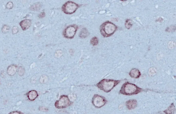
Microscopy imaging reveals TDP-43 pathology, early motor changes, neurodegeneration, and mitochondrial alterations in a TDP-43ΔNLS ALS model.
Introduction
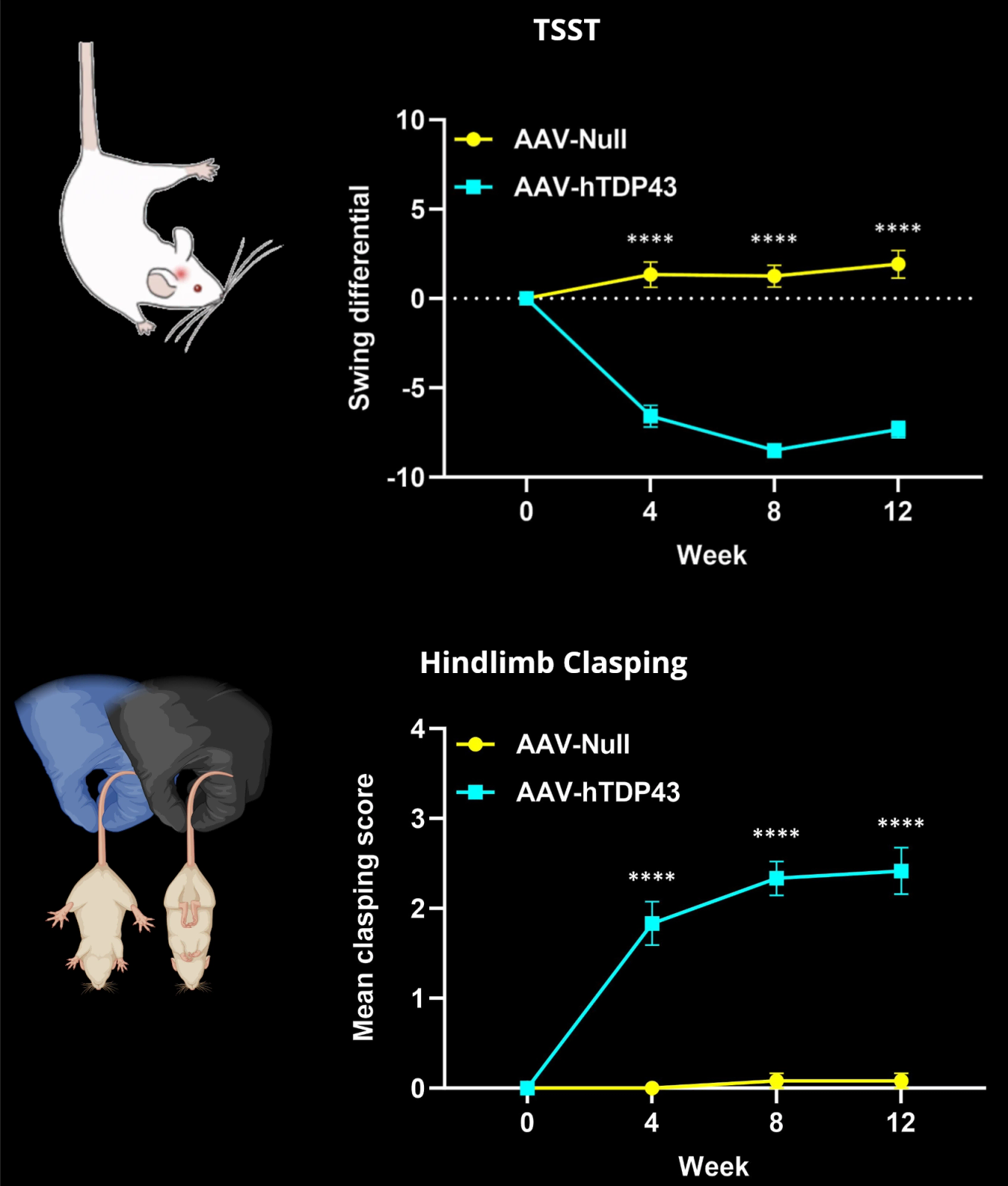
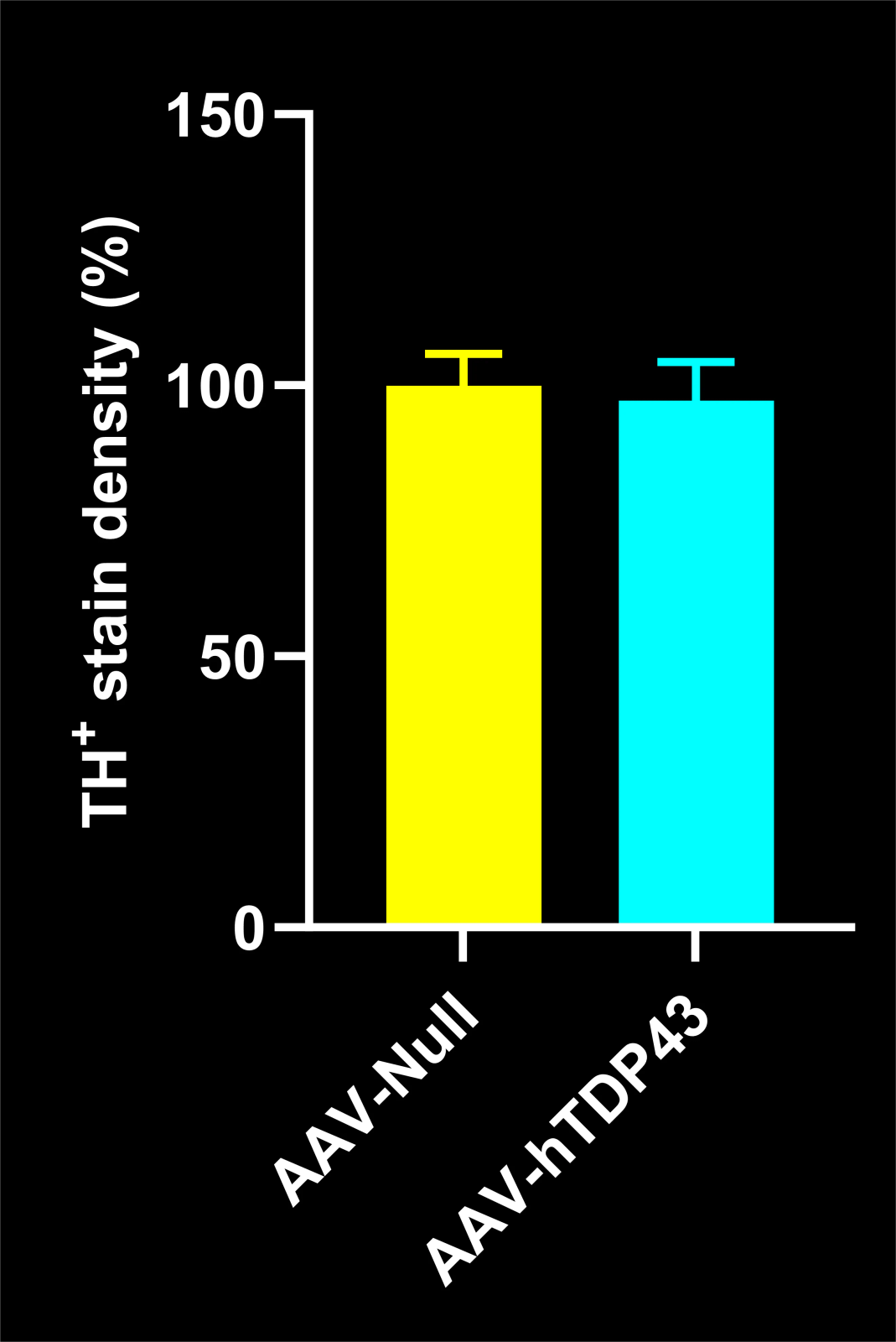
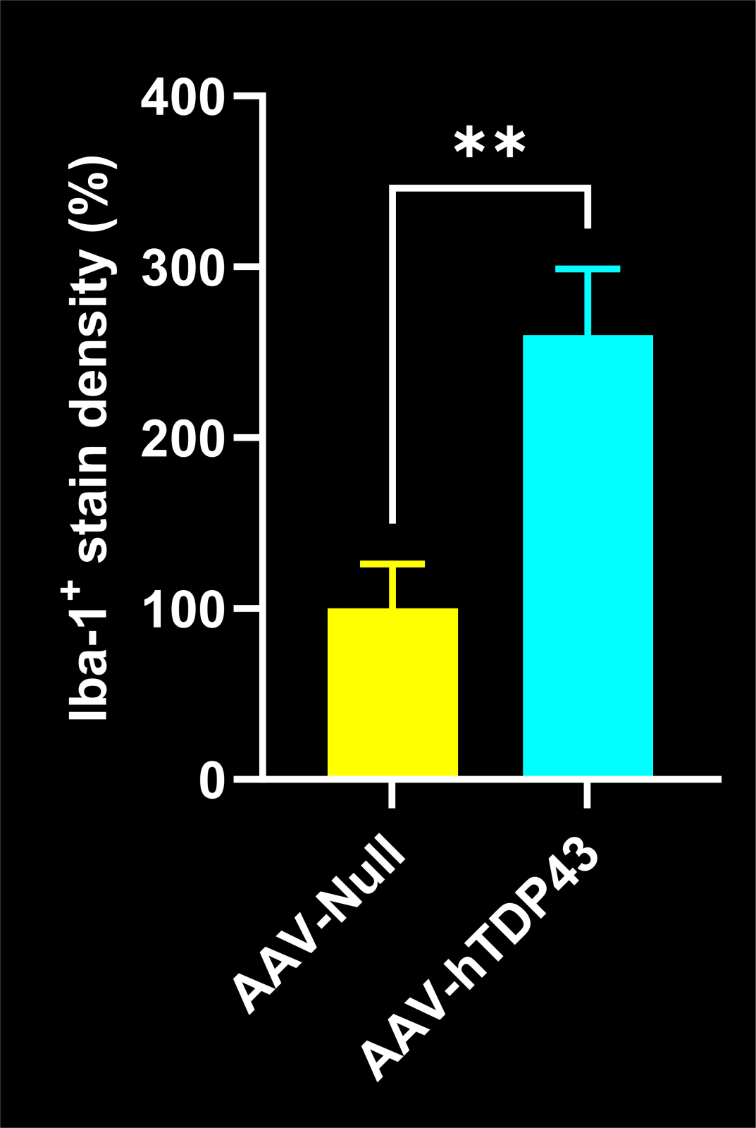
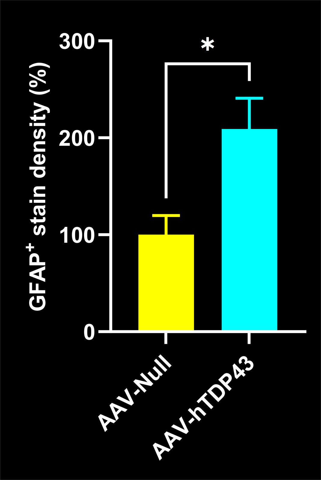
The AAV-TDP-43ΔNLS model expresses a cytoplasmically mislocalized form of human TDP-43, mimicking key pathological features seen in ALS. This includes phosphorylated TDP-43 (pTDP-43) aggregation, neuroinflammation, mitochondrial abnormalities, and motor deficits (Suk, 2020; Bright, 2021). Microscopy images presented here illustrate hallmark features of ALS-like pathology in this model.
Developed to provide flexibility in experimental design, this model enables controlled, region-specific targeting using AAV-based delivery. Moreover, this model can be rapidly established, facilitating faster study timelines for evaluating potential disease-modifying therapies.
This model is generated by unilaterally injecting 10-12-week-old C57BL/6 mice with either AAV-hTDP-43ΔNLS or AAV-Null (control) vectors into either the left motor cortex or the left substantia nigra pars compacta (SNc).
Image Interactive
This Story Panel in this interactive experience will guide you through our research project. You can also freely explore the high-resolution multiplex immunofluorescence tissue sections in the Image Viewer.


Key Assays used in this Research Project
Summary of some of the key behavioral and histological methods used to assess pathology in our AAV-TDP-43ΔNLS mouse model.
Behavioral Assays
Histological Assays
Would you like to contribute your own research to this Open Microscopy Initiative?
Related Content
Up-to-date information on ALS research and microscopy imaging.
A Guide to ALS Models for Drug Discovery
A Resource for the most effective use of research animal models (mouse & rat models) of Amyotrophic Lateral Sclerosis (ALS) for preclinical testing of therapeutics.
ALS Mouse Models & Spinal Motor Neurons
An overview of the involvement of spinal motor neurons in disease progression in mouse models of Amyotrophic Lateral Sclerosis (ALS).
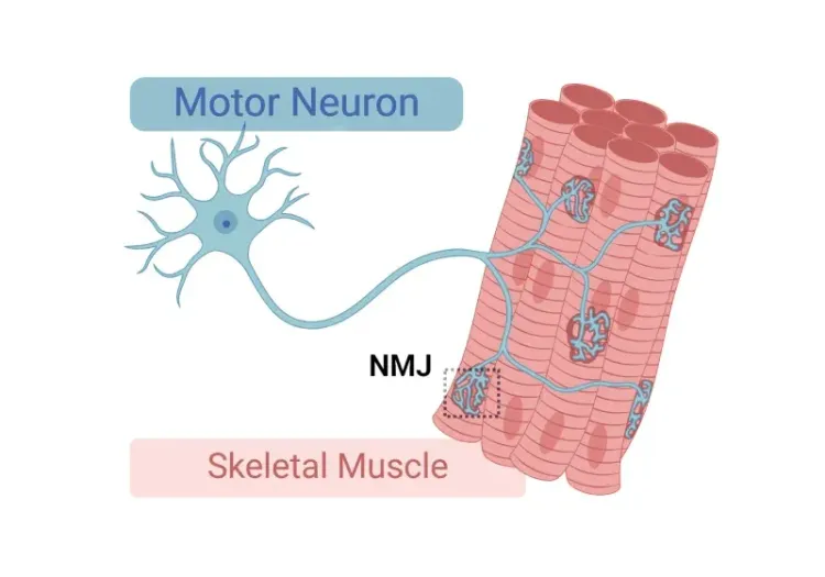
Neuromuscular Junction (NMJ) Morphology & ALS Models
Insights into neuromuscular junction (NMJ), its role in amyotrophic lateral sclerosis (ALS), and tools & methods used to study morphological changes in NMJs.