Characterization of a Novel AAV-hTau Mouse Model of Tauopathies with Parkinsonian Features
AAV-Tauマウスによるタウオパチーモデル
ヒトMAPT AAVベクターを注入して、タウ凝集体、神経炎症、神経変性、ドーパミン作動性神経の変性、運動障害を誘発します。
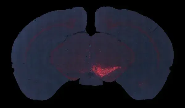
AAVタウオパチー(AAV-Tau)モデルの概要
進行性核上性麻痺および大脳基底核変性症のこのモデルでは、生後2~3ヶ月のC57BL/6マウスの黒質に、野生型ヒトタウ(MAPT)を過剰発現するAAVを片側から定位注入します。このマウスモデルは、ヒトのタウオパチーのいくつかの重要な特徴を再現しており、以下が含まれます。
- 黒質緻密部のドーパミン作動性神経細胞の減少
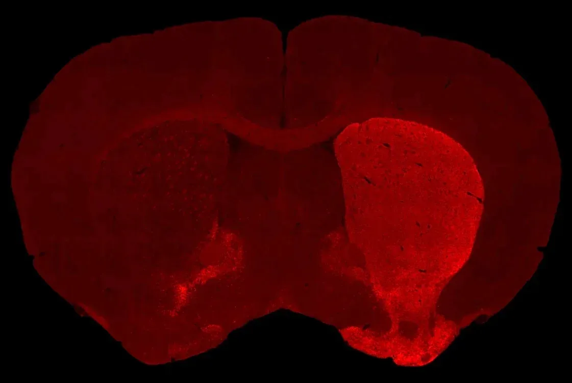
- 同側の線条体のドーパミン作動性神経の脱神経
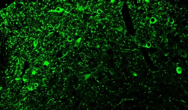
- 細胞体および神経突起におけるリン酸化タウの凝集体
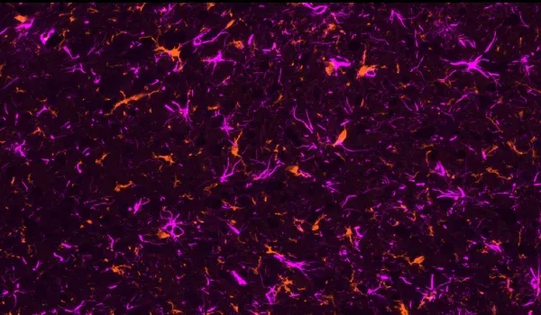
- 活性化されたミクログリア
- 反応性アストロサイト
- 運動機能障害
- 生体MRIスキャンで測定した脳萎縮(黒質、中脳、尾状核)
AAV-Tauモデルの生成
モデル生成の一般的なスキームは次のとおりです。

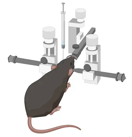
この特定のモデルでは、生後8~12週のC57BL/6マウスを使用します。次に、AAVベクターを黒質近辺に定位注入します。 高い精度と正確性を実現するために、自動マイクロインジェクター付きのデジタル定位装置を使用します。
このモデルを用いた研究は迅速に開始することができます。生体内試験は通常、約6週間続きます。そのため、特にアルツハイマー病やタウオパチーの従来のタウ遺伝子導入モデルと比較すると、比較的短期間で結果を得ることができます。
検証済みの対策
- 後肢把持テスト
- テールサスペンションのスイングテスト
- シリンダー試験
- ロータロッド試験
- MRI脳萎縮
- IHCおよび多重免疫蛍光法


注射したSNcにおけるAT8免疫蛍光染色。
注射したSNcにおけるIba-1とGFAPの二重免疫蛍光染色
同側半球におけるチロシン水酸化酵素染色の劇的な減少
注射されたSNcにおけるドーパミン作動性ニューロンの重度の消失
モデルの特性評価
以下のインタラクティブなプレゼンテーションでは、生体内データや多重免疫蛍光組織切片の全体像の高解像度画像など、当社のAAV-Tauマウスモデルの特性評価についてご覧いただけます。
左側のパネルを使って、この「イメージストーリー」を簡単にナビゲートすることができます。
高解像度の顕微鏡画像は、マウスの左ボタンで移動できます。 マウスまたはトラックパッド(上/下)または左上隅の + および - ボタンを使用して、拡大/縮小が可能です 。右上隅のコントロールパネル では、チャンネルとセグメンテーションの切り替え(オン/オフ)、色の変更、画像設定の調整が可能です。
最高のインタラクティブ体験 をお楽しみいただくには、フルスクリーンモード のご利用をお勧めします。
1/14
Tauopathies, such as Progressive Supranuclear Palsy and Corticobasal Degeneration, are rare diseases with prominent Parkinsonian features, including motor symptoms such as postural instability, vertical gaze palsy, rigidity, slowed movement (bradykinesia), muscle contractions (dystonia), and sudden jerks (myoclonus). Furthermore, individuals may face difficulties with speech and swallowing, cognitive decline, and loss of sensory perception at the cortical level. These neurodegenerative diseases are often rapidly progressing and pathologically characterized by phosphorylated tau inclusions in neurons and glia.
A significant problem for the development of disease-modifying therapeutics for tauopathies is the lack of animal models that recapitulate the human disease. To address this issue, Biospective has developed and characterized an adeno-associated virus (AAV) vector-induced mouse model that is well-suited for preclinical therapeutic efficacy studies for Progressive Supranuclear Palsy and Corticobasal Degeneration.
This Interactive Presentation illustrates some of the key motor function, neuroimaging, and pathologic features of Biospective's AAV human tau model of tauopathies with Parkinsonian features.
This model was generated by injecting 2 month-old C57BL/6 mice with AAV-hTau (wild-type 2N4R human tau) or AAV-null (control) vectors unilaterally into the left substantia nigra pars compacta (SNc) using a digital stereotaxic device with an automated microinjector.
Coronal Atlas View of SNc Injection Site
Multiplex immunofluorescence (mIF) images were generated by immunostaining for phosphorylated Tau (AT8), GFAP, Iba-1, Tyrosine Hydroxylase, Dopaminergic Nuclei, and counterstained with the DAPI nuclear stain. Tissue sections were digitized using a high-throughput slide scanner and were processed using Biospective's PERMITSTM software platform.
To navigate though this Image Story, you can use the arrows and/or the Table of Contents icon in the upper right corner of this panel.
You can also interact with the microscopy image in the viewer on the right at any time to further explore this high-resolution data.
Phosphorylated Human Tau Pathology
This microscopy image shows AT8 immunostaining for pTau. The ipsilateral (left hemisphere) midbrain shows extensive staining in the vicinity of the SNc and slightly beyond. For anatomical reference, an illustration with atlas labels for this brain level is provided below.
Coronal Mouse Brain Section (Bregma -3.2) with Neuroanatomy Labels
Tau Pathology in Neuronal Cell Bodies & Processes
This high magnification image shows extensive pTau staining in both the soma and processes of neurons in the SNc.
Neurodegeneration in the Substantia Nigra
As can be seen in this microscopy image, there is substantial loss of TH-positive dopaminergic neurons in the ipsilateral SNc compared to the contralateral hemisphere. For reference, an illustration with atlas labels for this brain level is provided below.
Coronal Mouse Brain Section (Bregma -3.2) with Neuroanatomy Labels
Using our PERMITSTM quantitative analysis software, we have quantified the TH staining in the SNc. The plot below shows a highly significant reduction in the ipsilateral hemisphere of the AAV-Tau compared to the AAV-null (control) mice.
TH stain density for AAV-Tau compared to AAV-null (control) injections; mean ± SEM, t-test, **** p<0.0001.
Brain Atrophy in the SNc and Midbrain
Regional brain atrophy is a key feature of tauopathies. Magnetic Resonance Imaging (MRI) is clinically used for non-invasive neuroimaging of Progressive Supranuclear Palsy (see our Resource) and Corticobasal Degeneration (see our Resource). Our team at Biospective has investigated the spatiotemporal pattern of brain atrophy in tauopathies (see MRI Measures of Disease Progression for Progressive Supranuclear Palsy Clinical Trials and MRI & Corticobasal Degeneration). We have found significant atrophy in multiple brain areas, including the midbrain and striatum in both diseases.
Given that MRI is a “translational biomarker”, we have acquired high-resolution in vivo anatomical 3D MR images from the AAV-hTau and AAV-null (control) mice using a 7T preclinical MRI scanner. We performed fully-automated image processing using our proprietary NIGHTWINGTM software and found highly significant brain atrophy in the SNc and midbrain. This data corresponds nicely to the loss of TH-positive neurons seen in the microscopy image.
Anatomical MRI with segmented SNc and midbrain, as well as plots of relative difference between ipsilateral and contralateral hemispheres for AAV-Tau compared to AAV-null (control) injections; mean ± SEM, t-test, **** p<0.0001.
Dopaminergic Neurons in the Contralateral SNc
This microscopy image shows the contralateral (right hemisphere) SNc which demonstrates TH-positive cell bodies and processes in red. The nuclei of the dopaminergic neurons are shown in blue.
Loss of Dopaminergic Neurons in the Ipsilateral SNc
This microscopy image shows the ipsilateral (left hemisphere) SNc which demonstrates a profound reduction of TH-positive cell bodies and processes (in red) compared to the contralateral hemisphere. The dopaminergic neuron nuclei are shown in blue.
Neurodegeneration in the Caudate-Putamen & Dopaminergic Motor Deficits
This microscopy image shows severe dopaminergic denervation of the ipsilateral (left hemisphere) caudate-putamen (loss of TH-positive terminals). For reference, an illustration with atlas labels for this approximate brain level is provided below.
Coronal Mouse Brain Section (Bregma +0.86) with Neuroanatomy Labels
Using our PERMITSTM quantitative analysis software, we have quantified the TH staining in the Caudate-Putamen. The plot below shows a highly significant reduction in the ipsilateral hemisphere.
TH stain density for AAV-Tau compared to AAV-null (control) injections; mean ± SEM, t-test, **** p<0.0001.
This loss of dopaminergic innervation corresponds well with unilateral motor deficits in these mice, including a highly significant increase in use of the ipsilateral paw during the Cylinder Test, decreased latency to fall in the Rotarod Test, increased swings to the contralateral side in the Tail Suspension Swing Test (TSST), and increased Hindlimb Clasping.
Cylinder Test, Rotarod Test, Tail Swing Suspension Test (TSST), and Hindlimb Clasping data for AAV-Tau compared to AAV-null (control) injections; mean ± SEM, t-test, **** p<0.0001.
Loss of Dopaminergic Terminals in the Ipsilateral Caudate-Putamen
This high magnification view shows the severe extent of loss of dopaminergic (TH-positive) terminals in the ipsilateral striatum. There are some remaining (albeit dystrophic) axons present.
We have also identified significant brain atrophy in the caudate-putamen on MRI scans, which aligns well with our analysis of human MRI data from Progressive Supranuclear Palsy and Corticobasal Degeneration populations. This data supports the “translatability” of this tauopathy model.
Anatomical MRI with segmented striatum, as well as plot of relative difference between ipsilateral and contralateral striatum. ****p<0.0001.
Microgliosis in Response to Human 2N4R Tau Expression
In this low magnification image, one can readily appreciate the higher density of Iba-1-positive microglia in the ipsilateral (left) hemisphere (indicated by the box) relative to the contralateral hemisphere.
The plot below shows the Iba-1 stain density in the SNc.
Iba-1 stain density for AAV-Tau compared to AAV-null (control) injections; mean ± SEM, t-test, *** p<0.001.
We have performed a morphological analysis of microglia using a novel computer vision & machine learning approach developed by our team. This fully-automated algorithm classifies non-activated (ramified) and activated (non-ramified) microglia.
The plot below shows the microglial activation in the SNc, with highly significant increased microglial activation in the AAV-Tau mice.
Microglial activation for AAV-Tau compared to AAV-null (control) injections; mean ± SEM, t-test, *** p<0.0001.
Iba-1 Staining in Proximity to Phosphorylated Tau
This high magnification view shows the increased density of Iba-1-stained microglia in areas with phosphorylated tau aggregates.
Astrogliosis & Human Tau Pathology
This low magnification microscopy image show a higher density of GFAP-positive astrocytes in the ipsilateral hemisphere (indicated by the box). The plot below shows the GFAP stain density in the SNc.
GFAP stain density for AAV-Tau compared to AAV-null (control) injections; mean ± SEM, t-test, **** p<0.0001.
GFAP Staining in Proximity to p-Tau
This high magnification view shows the increased density of GFAP-stained astrocytes in areas with phosphorylated Tau aggregates.
Summary
This novel mouse model of tauopathies with Parkinsonian features recapitulates many of the hallmark features of Progressive Supranuclear Palsy and Corticobasal Degeneration, including the development of asymmetric motor dysfunction and (due to unilateral injection), and associated loss of TH+ SNc neurons and striatal TH expression.
AAV-hTau regionally results in highly significant brain atrophy, elevated microglial density and activation levels, and increased astrocyte density and hypertrophy. Further studies are planned to continue to investigate the pathologic changes in this model.
This inducible and rapidly progressing mouse model is well-suited for drug discovery with quantitative in vivo and ex vivo readouts, and possesses distinct advantages over existing transgenic models as a screening method for novel treatment options targeting tau-related pathology.
Please feel free to further explore the microscopy image in the viewer.
We would be happy to discuss this model and our characterization if you would like to Contact Us.
当社のAAV-Tauマウスモデルの特性評価、検証済みの測定方法、および前臨床神経科学CROサービスについて、さらに詳しく知ることができます。
アルツハイマー病とタウオパチーのモデルをもっと見る
MRIによる脳萎縮は、翻訳バイオマーカーとして使用できるでしょうか?
MRIで測定される局所脳萎縮は、動物モデル研究とヒトの臨床試験のギャップを埋めるトランスレーショナル・イメージング・バイオマーカーです。 私たちは、進行性核上性麻痺、皮質基底核変性症、および前頭側頭型認知症におけるMRIバイオマーカーの使用について説明するリソースおよびイノベーションプレゼンテーションをいくつか発表しています。
脳の萎縮は主にタウタンパク質またはアミロイドβによって引き起こされるのでしょうか?
Biospectiveの私たちのグループは、この重要なテーマを徹底的に研究してきました。最近、私たちは学術誌に論文を発表しました。アルツハイマー病と認知症:
また、このトピックに関するイノベーションプレゼンテーションも公開しています。
このタウオパチーモデルにおける尾状核・被殻のドーパミン作動性神経の変性を定量的に評価することはできますか?
はい。弊社では、デジタル化した組織切片から定量的測定値を導き出すために、独自開発のPERMITS™ソフトウェアを使用しています。線条体におけるドーパミン作動性終末の損失を評価するために、チロシン水酸化酵素(TH)に対する免疫染色を行い、TH陽性プロセスを定量化します。
「テールサスペンションスイングテスト」とは何ですか?
尾懸垂振り子テストは、実施が容易な試験であり、齧歯類の運動の非対称性を評価するために使用されます。この試験では、動物を尾で地面から約5cmの高さに吊り下げ、頭部を垂直軸の外側に振り向ける回数と方向を数えます。
この行動テストは、初期の運動障害の検出、神経変性の程度評価、治療による神経保護効果の特性評価に使用されます。
AAV-Tauモデルにおける「反応性」アストロサイトの定量化はどのように行いますか?
Biospectiveの当社チームは、免疫組織化学および多重免疫蛍光画像における形態学に基づいて反応性アストロサイトを定量化する先進的なコンピュータービジョンおよび機械学習アルゴリズムを開発しました。詳細は、当社のプレゼンテーション「アストロサイトおよびアミロイドβのアルツハイマー病マウスモデル」をご覧ください。
「アデノ随伴ウイルス(AAV)」ベクターとは何ですか?
組み換えアデノ随伴ウイルス(rAAV)は、研究および臨床応用に広く使用されている遺伝子導入ツールです。AAVは、4.8kbのDNA分子を持つ一本鎖DNAウイルスです。遺伝子治療や体細胞遺伝子導入では、ウイルスDNAは目的の遺伝子(GOI)を含む設計されたDNAに置き換えられます。
関連コンテンツ
アルツハイマー病およびタウオパチーに関する最新情報と、動物モデルにおける治療薬の評価のための翻訳バイオマーカーの使用に関するベストプラクティス。
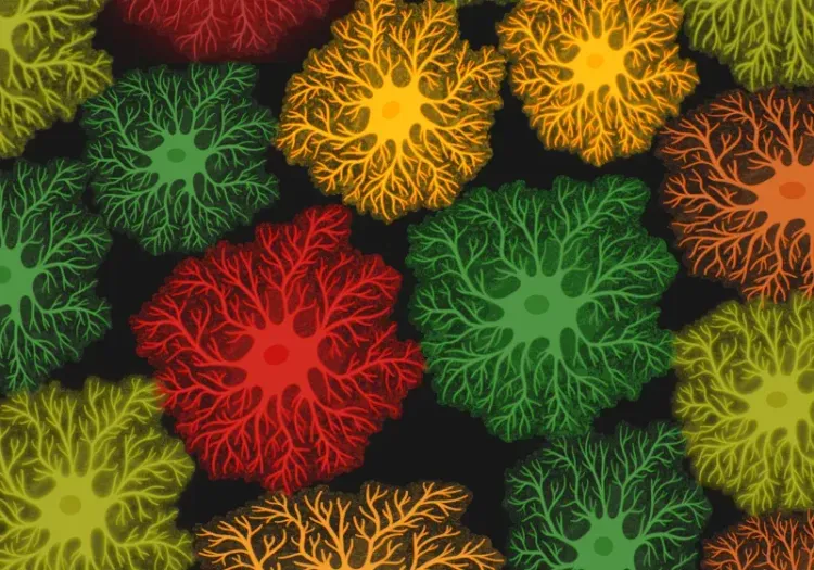
アルツハイマー病におけるアストロサイトの形態
アストロサイトの形態解析の概要と、神経変性疾患の研究および創薬・薬剤開発への応用。
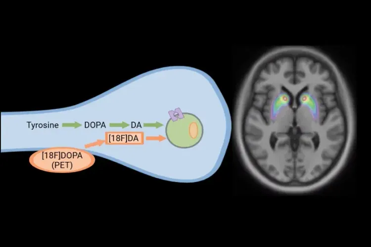
[18F]DOPA PETによるパーキンソン病臨床試験
パーキンソン病および運動障害の臨床試験における、[18F]DOPA PETによる疾患進行のモニタリングと治療介入への反応の評価方法。