AAV 타우병증(AAV-Tau) 모델 개요
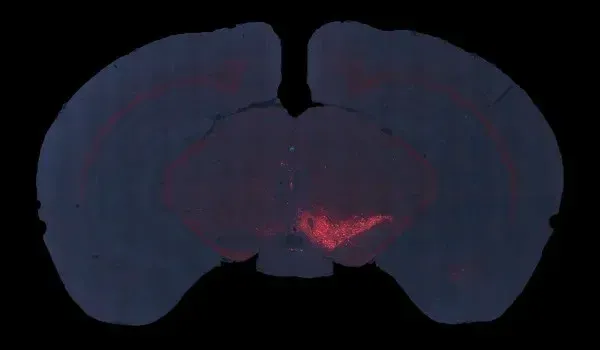
이 진행성 핵상마비 및 피질-기저변성 모델의 경우, 생쥐의 약 2-3개월령에 C57BL/6 마우스의 흑질에 야생형 인간 타우(MAPT)를 과발현하는 AAV를 일측성 정위 주사합니다. 이 마우스 모델은 다음과 같은 인간 타우병의 몇 가지 주요 특징을 재현합니다.
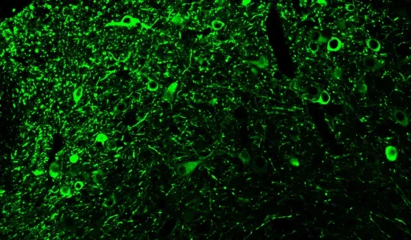
- 흑질 소구체의 도파민성 뉴런 감소
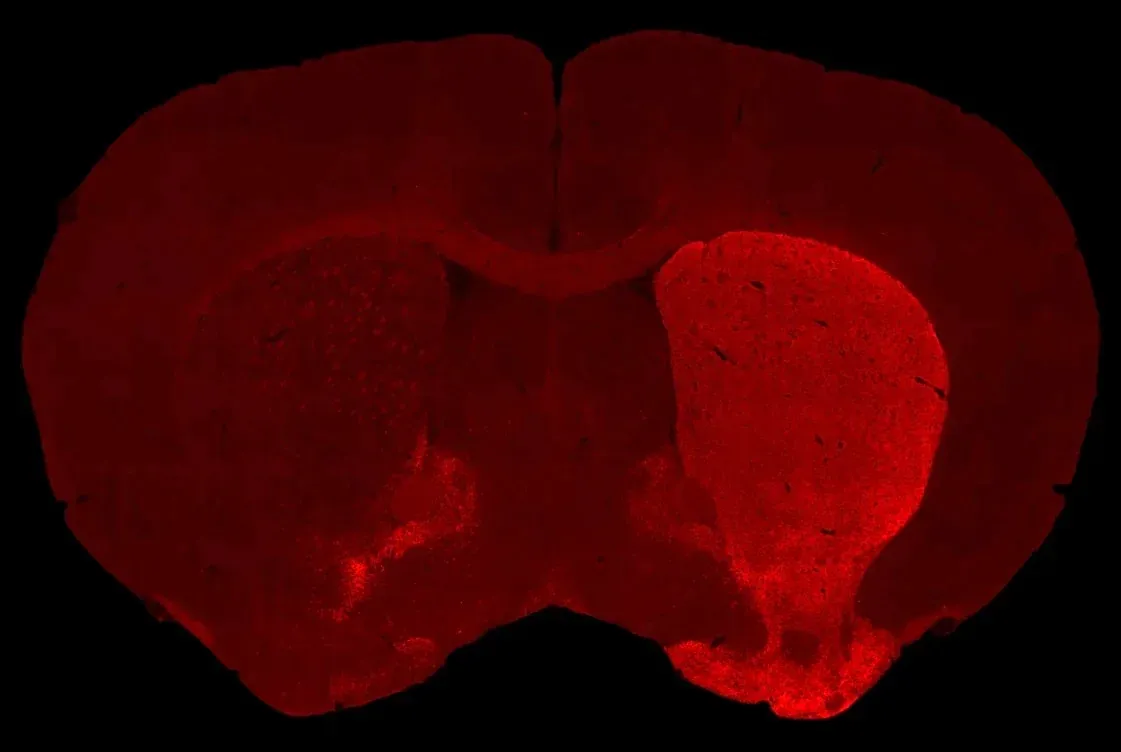
- 동측 선조체의 도파민 신경절 절제
- 세포체와 신경돌기에 있는 인산화 타우의 응집체
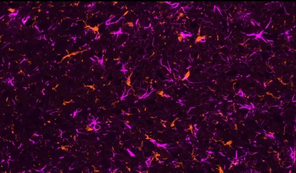
- 활성화된 미세아교세포
- 반응성 성상세포
- 운동 기능 장애
- 생체 내 MRI 스캔으로 측정한 뇌 위축(흑질, 중뇌, 후두핵)
AAV-Tau 모델 생성
모델 생성을 위한 일반적인 스키마는 다음과 같습니다.
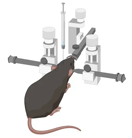
이 특정 모델의 경우, 생후 8-12주령의 C57BL/6 마우스를 사용합니다. 그런 다음, AAV 벡터를 흑질 부근에 정위 주사합니다. 높은 정확성과 정밀성을 위해 자동 마이크로 주사기가 장착된 디지털 정위 장치를 사용합니다.
이 모델을 이용한 연구는 신속하게 시작할 수 있습니다. 생체 내 단계는 일반적으로 약 6주 동안 지속됩니다. 따라서, 특히 기존의 알츠하이머병 및 타우병에 대한 타우 트랜스제닉 모델과 비교할 때, 비교적 짧은 시간 내에 결과를 얻을 수 있습니다.
검증된 조치
- 뒷다리 쥐기 테스트
- 테일 서스펜션 스윙 테스트
- 실린더 테스트
- 로타로드 테스트
- MRI 뇌 위축
- IHC & 멀티플렉스 면역 형광법
이 모델이 인간 타우병에 적용될 수 있는지에 대해 자세히 알아보세요.
모델 특성화
아래의 대화형 프레젠테이션을 통해 생체 내 데이터와 전체 다중 면역형광 조직 섹션의 고해상도 이미지를 포함하여 AAV-Tau 마우스 모델의 특징을 살펴볼 수 있습니다.
왼쪽 패널을 사용하여 이 "이미지 스토리"를 간단하게 탐색할 수 있습니다.
왼쪽 마우스 버튼을 사용하여 고해상도 현미경 이미지를 이동할 수 있습니다. 마우스/트랙패드(위/아래) 또는 왼쪽 상단 모서리에 있는 + 및 - 버튼 을 사용하여 확대 및 축소할 수 있습니다 . 오른쪽 상단 모서리에 있는 제어판에서 채널 및 세분화에 대한 이미지 설정을 토글(켜기/끄기), 색상 변경 및 조정할 수 있습니다.
최상의 상호작용 경험을 위해 전체 화면 모드를 사용하는 것이 좋습니다.
AAV-Tau 마우스 모델의 특성, 검증된 측정 방법, 전임상 신경과학 CRO 서비스에 대해 자세히 알아보세요.
타우병 모델을 더 알아보세요
관련 콘텐츠
알츠하이머병 및 타우병에 대한 최신 정보와 동물 모델에서 치료제 평가를 위한 번역적 바이오마커 사용과 관련된 모범 사례.
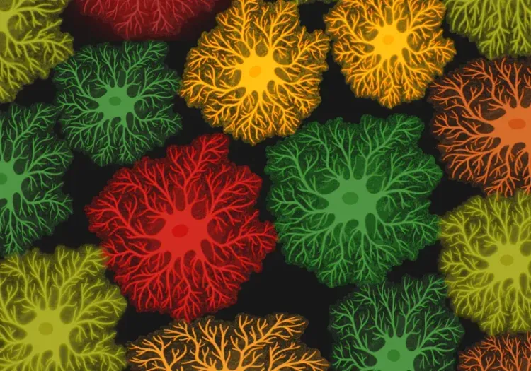
알츠하이머병의 성상교세포 형태
성상교세포 형태학적 분석의 개요와 신경 퇴행성 질환 연구 및 신약 개발에 대한 응용.
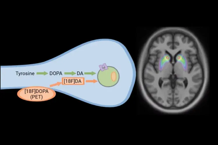
파킨슨병 임상시험에서 [18F]DOPA PET
파킨슨병 및 운동장애 임상시험에서 질병의 진행과 치료적 개입에 대한 반응을 모니터링하기 위해 [18F]DOPA PET가 어떻게 사용되는지 알아보세요.