Therapeutic Areas
We have extensive experience with quantitative imaging biomarkers for a range of neurological diseases. We use these robust imaging measures to assess study eligibility and therapeutic efficacy in early to late phase clinical trials.

Imaging Modalities
Our 21 CFR Part 11 & GCP compliant, validated, quantitative image processing and analysis software platform is purpose-built for clinical trials. Our expert team of imaging scientists and software developers leverage cutting-edge methods, including machine learning and deep learning algorithms, for robust fully-automated image processing & analysis of multi-modality MRI, PET, and SPECT data.
- Volumetric MRI
- Amyloid PET
- Tau PET
- FDG PET
- Dopaminergic Imaging
- fMRI
- Diffusion MRI
- ASL MRI
- Neuromelanin MRI
- SWI
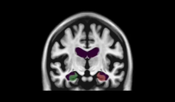
Volumetric MRI
Using 3D T1-weighted MRI scans, we can quantitatively assess changes in regional brain volumes and cortical thickness. The specific measures can be configured for a particular therapeutic area.
For example, typical measures for Alzheimer’s disease include hippocampus, lateral ventricle, and whole brain volumes, as well as cortical thickness. For Multiple System Atrophy, we can evaluate the volumes of the caudate, pallidum, putamen, cerebellum, and brainstem.
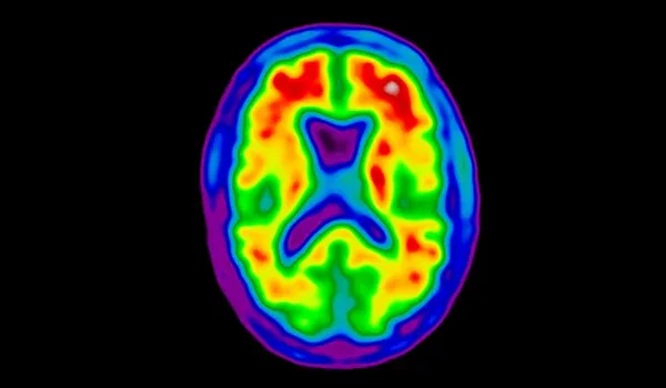
Amyloid PET
Amyloid PET is a non-invasive diagnostic imaging technique used to detect beta-amyloid plaques in the brain. We have extensive experience processing and analyzing Amyloid PET images derived from multiple different beta-amyloid radiotracers used in multicenter clinical trials.
We can generate SUVR, Centiloid, and other sensitive measures. We can also perform nuclear medicine reads for study eligibility and subject enrichment.
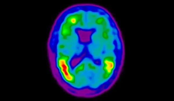
Tau PET
Tau proteins are linked to numerous neurodegenerative conditions, such as Alzheimer's disease, specific types of frontotemporal dementia (FTD), and other tauopathies. Tau PET imaging is instrumental in both diagnosing and following the progression of these diseases. Additionally, it plays a pivotal role in monitoring the effectiveness of therapeutic interventions during clinical trials.
We use can quantitatively assess regional tau burden and the spatial extent of tau pathology using our fully-automated image processing & analysis techniques.
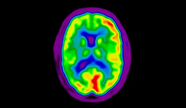
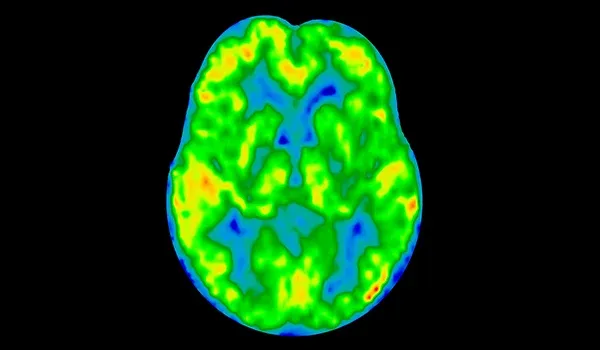
FDG PET
FDG (fluorodeoxyglucose) PET imaging plays a crucial role in understanding brain function and monitoring changes in disease progression and response to therapeutic intervention. In many neurodegenerative diseases, including Alzheimer’s disease and Parkinson’s disease, there are characteristic spatiotemporal patterns of decreased and increased glucose metabolism in the brain. FDG PET imaging can be implemented in multicenter clinical trials, and we can measure regional SUVR changes.

Dopaminergic Imaging
SPECT and PET tracers that visualize dopaminergic terminals, including dopamine transporter (DaT) ligands (e.g. DaTscan), VMAT2 receptor ligands (e.g. [18F]AV-133), and [18F]fluorodopa (FDOPA), can be used to non-invasively evaluate dopaminergic degeneration. Dopaminergic SPECT or PET imaging is widely used in clinical trials of Parkinson’s disease.
We have developed fully-automated SBR and SUVR measures to assess change in the dopamine system. We can also perform nuclear medicine reads of these images for trial inclusion and subject enrichment.
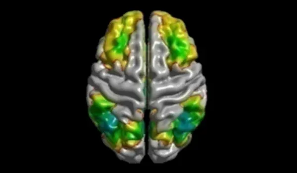
fMRI
We have implemented automated pre-processing algorithms and sophisticated statistical analysis for functional MRI (fMRI) scans. fMRI maps brain activity by detecting changes in cerebral blood flow and oxygenation levels. fMRI provides insights into which areas of the brain are activated during various tasks or while the brain is at rest.
fMRI is increasingly being utilized in clinical trials, especially in neurodegenerative diseases, to evaluate the effects of therapeutic intervention on brain function.
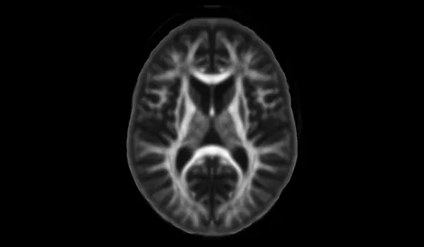
Diffusion MRI
Diffusion MRI is used to examine microstructural changes in white matter of the brain and spinal cord. Simple diffusivity measures are used in diseases such as Multiple System Atrophy. Diffusion Tensor Imaging (DTI) is a commonly used model which generates several metrics (FA, MD, AD, RD). We can also estimate free water from DTI scans. More sophisticated models, such as NODDI, can also be used to estimate intracellular and extracellular fractions. These measures can be useful in MS, neurodegenerative diseases, and genetic & rare diseases.
ASL MRI
Arterial Spin Labeling (ASL) perfusion MRI is used to estimate cerebral blood flow (CBF) in the brain. ASL utilizes magnetically-labeled arterial blood water as an endogenous tracer to provide quantitative information about brain perfusion without the need for contrast agent injection.
Perfusion is tightly coupled with glucose metabolism (typically measured by FDG PET), and cerebral hypoperfusion & hypometabolism are well-established biomarkers for several neurodegenerative diseases.
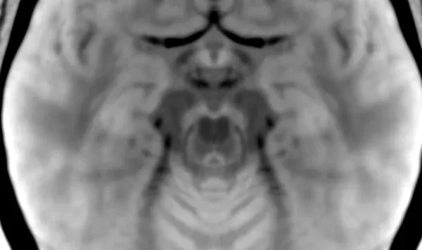
Neuromelanin MRI
Neuromelanin MRI (NM-MRI) is a cutting-edge imaging technique used to visualize neuromelanin-containing structures within the brain, particularly the substantia nigra and locus coeruleus.
We have developed a standardized image acquisition protocol for high-resolution 3D NM-MRI data that works across MRI scanner models. We can automatically generate several quantitative measures derived from NM-MRI data for eligibility and efficacy assessments in clinical trials of Parkinson’s disease.
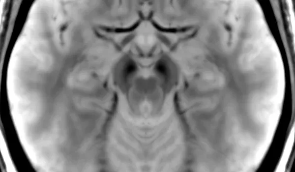
SWI
Susceptibility-weighted MRI (SWI) is an advanced imaging technique that enhances the visualization of brain structures and abnormalities (e.g. microhemorrhages) by exploiting differences in the magnetic susceptibility of tissues.
SWI can be particularly useful for evaluating iron-related changes in the substantia nigra in Parkinson’s disease. We have implemented a robust protocol for simultaneous acquisition of 3D SWI and neuromelanin MRI (NM-MRI) for Parkinson’s disease clinical trials.
Learn more about our Image Processing & Analysis services for clinical trials.